Increased expression of hypoxia-induced factor 1α mRNA and its related genes in myeloid blood cells from critically ill COVID-19 patients
←
→
Transcripción del contenido de la página
Si su navegador no muestra la página correctamente, lea el contenido de la página a continuación
Increased expression of hypoxia-induced factor 1α
mRNA and its related genes in myeloid blood cells
from critically ill COVID-19 patients
Keiko Taniguchi-Ponciano
 Unidad de Investigación Médica en Enfermedades Endocrinas, UMAE Hospital de Especialidades, Centro
Medico Nacional Siglo XXI, Instituto Mexicano del Seguro Social.
Eduardo Vadillo
 Unidad de Investigación Médica en Enfermedades Oncológicas, UMAE Hospital de Oncología, Instituto
Mexicano del Seguro Social.
Héctor Mayani
 Unidad de Investigación Médica en Enfermedades Oncológicas, UMAE Hospital de Oncología, Instituto
Mexicano del Seguro Social.
César Raúl Gonzales-Bonilla
 Titular, Coordinación de Investigación en Salud, Instituto Mexicano del Seguro Social.
Javier Torres
 Unidad de Investigación Médica en Enfermedades Infecciosas y Parasitarias, UMAE Hospital de
Pediatría, Instituto Mexicano del Seguro Social.
Abraham Majluf
 Unidad de Investigación Médica en trombosis, hemostasia y aterogénesis, Instituto Mexicano del
Seguro Social.
Guillermo Flores-Padilla
  Servicio de Medicina Interna, UMAE Hospital de Especialidades, Centro Medico Nacional Siglo XXI,
Instituto Mexicano del Seguro Social.
Niels Wacher-Rodarte
 Unidad de Investigación Médica en Epidemiología Clínica, UMAE Hospital de Especialidades, Centro
Medico Nacional Siglo XXI, Instituto Mexicano del Seguro Social.
Juan Carlos Galan
  Servicio de Medicina Interna, UMAE Hospital de Especialidades, Centro Medico Nacional Siglo XXI,
Instituto Mexicano del Seguro Social.
Eduardo Ferat-Osorio
 División de Investigación en Salud, UMAE Hospital de Especialidades, Centro Medico Nacional Siglo
XXI, Instituto Mexicano del Seguro Social.
Francisco Blanco-Favela

                                                Page 1/14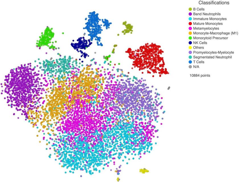
Unidad de Investigación Médica en Inmunología, UMAE Hospital de Pediatría, Centro Medico Nacional
Siglo XXI, Instituto Mexicano del Seguro Social.
Constantino Lopez-Macias
Unidad de Investigación Médica en Inmunoquímica, UMAE Hospital de Especialidades, Centro Medico
Nacional Siglo XXI, Instituto Mexicano del Seguro Social.
Aldo Ferreira-Hermosillo
Unidad de Investigación Médica en Enfermedades Endocrinas, UMAE Hospital de Especialidades, Centro
Medico Nacional Siglo XXI, Instituto Mexicano del Seguro Social.
Claudia Ramirez-Renteria
Unidad de Investigación Médica en Enfermedades Endocrinas, UMAE Hospital de Especialidades, Centro
Medico Nacional Siglo XXI, Instituto Mexicano del Seguro Social.
Eduardo Peña-Martínez
Unidad de Investigación Médica en Enfermedades Endocrinas, UMAE Hospital de Especialidades, Centro
Medico Nacional Siglo XXI, Instituto Mexicano del Seguro Social.
Gloria Silva-Román
Unidad de Investigación Médica en Enfermedades Endocrinas, UMAE Hospital de Especialidades, Centro
Medico Nacional Siglo XXI, Instituto Mexicano del Seguro Social.
Sandra Vela-Patiño
Unidad de Investigación Médica en Enfermedades Endocrinas, UMAE Hospital de Especialidades, Centro
Medico Nacional Siglo XXI, Instituto Mexicano del Seguro Social.
Carlos Mata-Lozano
Unidad de Investigación Médica en Enfermedades Endocrinas, UMAE Hospital de Especialidades, Centro
Medico Nacional Siglo XXI, Instituto Mexicano del Seguro Social.
Roberto Carvente-Garcia
Unidad de Investigación Médica en Enfermedades Endocrinas, UMAE Hospital de Especialidades, Centro
Medico Nacional Siglo XXI, Instituto Mexicano del Seguro Social.
Lourdes Basurto-Acevedo
Unidad de Investigación Médica en Enfermedades Endocrinas, UMAE Hospital de Especialidades, Centro
Medico Nacional Siglo XXI, Instituto Mexicano del Seguro Social.
Renata Saucedo
Unidad de Investigación Médica en Enfermedades Endocrinas, UMAE Hospital de Especialidades, Centro
Medico Nacional Siglo XXI, Instituto Mexicano del Seguro Social.
Patricia Piña-Sanchez
Unidad de Investigación Médica en Enfermedades Oncológicas, UMAE Hospital de Oncología, Instituto
Mexicano del Seguro Social.
María Antonieta Chavez-Gonzalez
Unidad de Investigación Médica en Enfermedades Oncológicas, UMAE Hospital de Oncología, Instituto
Mexicano del Seguro Social.
Daniel Marrero-Rodríguez ( dan.mar57@gmail.com )
Page 2/14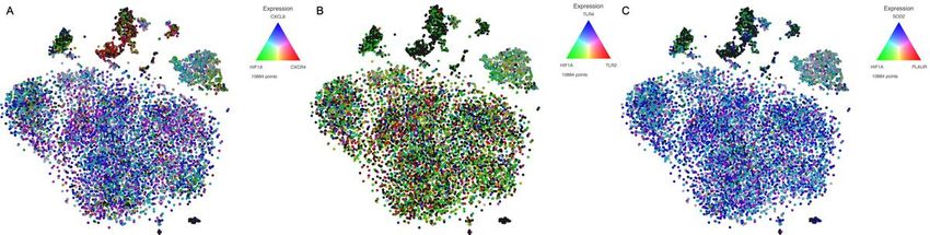
Unidad de Investigación Médica en Enfermedades Endocrinas, UMAE Hospital de Especialidades, Centro
Medico Nacional Siglo XXI, Instituto Mexicano del Seguro Social.
Moisés Mercado ( moises.mercado@endocrinologia.org.mx )
Unidad de Investigación Médica en Enfermedades Endocrinas, UMAE Hospital de Especialidades, Centro
Medico Nacional Siglo XXI, Instituto Mexicano del Seguro Social.
Research Article
Keywords: SARS-Cov-2, COVID-19, critically-ill, scRNAseq, HIF1A, myeloid
Posted Date: July 17th, 2020
DOI: https://doi.org/10.21203/rs.3.rs-43390/v1
License: This work is licensed under a Creative Commons Attribution 4.0 International License.
Read Full License
Version of Record: A version of this preprint was published at Annals of Medicine on December 21st,
2020. See the published version at https://doi.org/10.1080/07853890.2020.1858234.
Page 3/14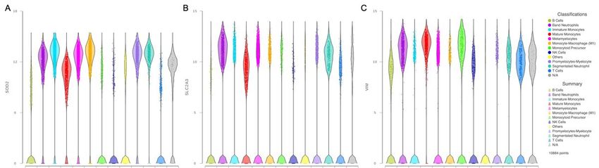
Abstract
Since its emergence, in December 2019, COVID-19 has resulted in more than 12 million people infected
and has killed more than 570000. Hypoxemia has been identified as one of the main clinical
manifestations of this disease, especially in severe cases. We have previously reported that in critically ill
COVID-19 patients there is a shift towards an immature myeloid profile in peripheral blood cells, including
band neutrophils, immature monocytes, metamyelocytes, monocyte-macrophages, monocytoid
precursors, and promyelocytes-myelocytes, which, together with mature monocytes and segmented
neutrophils, comprise the vast majority of blood cells in these patients. Such an immature myeloid profile
may be the result of a physiological response known as emergency myelopoiesis. In the present study, we
performed scRNAseq from leukocytes from five critically ill COVID-19 patients and characterized the
expression of hypoxia-inducible factor1α (HIF1α) mRNA and its transcriptionally regulated genes. HIF1α
is a master transcription factor involved in the cellular response to hypoxia. We herein report that these
cellular subsets express high levels of HIF1α mRNA and several of their transcriptional targets, including
those related to inflammation, such as CXCL8, CXCR1, CXCR2, and CXCR4; those potentially involved in
virus sensing, such as TLR2 and TLR4; and those related to metabolism, such as SLC2A3, PFKFB3, PGK1,
GAPDH and SOD2. The up-regulation and participation of HIF1α in relevant events such as inflammation,
immunometabolism, and TLR make it a potential molecular marker for COVID-19 severity and,
interestingly, could represent a potential target for molecular therapy.

Introduction
The coronavirus disease 2019 (COVID–19) epidemic caused by the severe acute respiratory syndrome
coronavirus 2 1 has rapidly developed into a devastating pandemic 1. As of today, the World Health
Organization has reported more than 12 million persons diagnosed with COVID–19 and over 570 000
deaths worldwide2. It is associated with significant mortality in high risk patients, with poor prognostic
features upon admission. The spectrum of the disease is broad, including pneumonia, sepsis, and acute
respiratory distress syndrome (ARDS) 3. Hypoxemia, defined as a decrease in the partial pressure of
oxygen is an ominous sign of COVID–19, and it is usually an indicator of disease severity 4,5. An oxygen
saturation above 90% is associated with better outcomes 6. Over 80% of COVID–19 patients in the
intensive care unit have severe hypoxemia 7. A kind of “silent hypoxia” in which COVID–19 patients
deteriorate rapidly without warning and develop respiratory failure has been described 8. Hypoxia
indicates an imbalance of oxygen delivery to tissues and leads to compromised function, which is
quantitatively related to organ, tissue and even cell type 9. The hypoxia-inducible factors (HIF) are
considered master regulators of oxygen homeostasis and are oxygen level sensitive 10. Currently, there is
scarce information regarding the expression of HIF in patients with severe COVID–19 and its potential
involvement in the immunopathogenesis of this condition. Therefore, in the present work we carried out
scRNAseq to identify the cell populations present in critically ill COVID–19 patients and to determine the
expression of hypoxia-induced factor 1α (HIF1α) and its related genes.

                                                   Page 4/14
Materials And Methods
Patients and tissue samples
Blood samples from five critically ill patients with COVID–19 were collected in EDTA-coated tubes.
Tissues were collected from patients diagnosed, treated and followed at the Medicina Interna department
of the Hospital de Especialidades, Centro Médico Nacional Siglo XXI of the Instituto Mexicano del Seguro
Social in April 2020. A family member of each participating patient signed an informed consent and the
study protocol was approved by the Comisión Nacional de Ética e Investigación Científica del Instituto
Mexicano del Seguro Social in accordance to the Helsinki declaration. SARS-CoV–2 infection was
corroborated by RT-qPCR at an official federal government reference laboratory.
Sample preparation, scRNAseq library generation and
sequencing
Peripheral blood from the five critical COVID–19 patients was collected in EDTA-coated tubes, and
immune cells were isolated according to standard centrifugation methods followed by red blood cell
lysis.
Chromium Next GEM Single Cell 3´ Reagent Kits v3.1 and protocol from 10X Genomics was followed as
recommended by manufacturer’s instructions. Briefly, immune cells were pooled in a single tube and cells
were diluted in 1x phosphate buffered saline (PBS) to 700–1200 cells per µl. Cell suspension was loaded
in Chromium Next GEM Chip G and sorted in the Chromium Controller from 10X Genomics. The Cell-Gel
Beads in Emulsion (GEMs) were then incubated to generate the barcoded cDNA. cDNA was cleaned using
Dynabeads and washed, followed by cDNA amplification and SPRIselection. The retrieved cDNA was
enzymatically fragmented, end-repaired, poly-A tailed and ligated. Size selection, adaptor ligation and
amplification were done. Sequencing was done using NextSeq 550 System High-Output Kit (300 cycles)
in NextSeq 500 system (Illumina) according to 10X Genomics specifications: Read 1 = 28 cycles, Read 2
= 91 cycles, Index 1 = 8 cycles. All quality control steps were carried out using 4200 TapeStation System
(Agilent) with High Sensitivity D1000 Screen Tape, whereas the concentration was calculated using Qubit
2.0 Fluorometer with Kit High Sensitivity assays.
scRNAseq bioinformatic analysis
Partek Flow software was used with scRNAseq toolbox. First the tags were trimmed and then the reads
were aligned using STAR 2.7.3a algorithm to human genome hg38. UMI’s were deduplicated and barcode
filtered. Following criteria were then applied to each cell, i.e., gene number between 200 and 6000, UMI
count above 300 and mitochondrial gene percentage below 20%. To quantify the transcriptome human
hg38 Ensembl transcripts release 99 was used. Counts per million, Add 1.0 Log 2.0 were the
normalization parameters. Healthy donors’ datasets were downloaded from 10X Genomics website and
Page 5/14analyzed using Loupe Browser from 10X Genomics. Data has been deposited in Sequence Read Archive
hosted by National Center for Biotechnology Information under accession number.
Markers used to circumscribe cell populations
Clusters were categorized by analyzing differentially expressed genes according to previously published
data obtained from human samples11–16
Dimensionality reduction and clustering
The filtered and normalized gene-barcode matrix was analyzed by principal components, then graph
based and t-distributed stochastic neighbor embedding (t-SNE) using default parameters was carried out.
Results
Immature myeloid cell populations in critically ill COVID–19
patients
We have previously reported that, as compared to healthy adults, lymphoid cell subsets, such as B and T
lymphocytes as well as NK cells, were present in low quantities in critically ill COVID–19 patients,
whereas cells of myeloid origin predominated. Interestingly, immature myeloid cell populations, such as
band neutrophils, metamyelocytes, promyelocytes-myelocytes, monocytoid precursor, and immature
monocytes prevailed. Mature lineages such as segmented neutrophils, mature monocytes and finally
monocyte-macrophages were also observed (Figure 1) 17.
HIF1α expression in leukocytes from critically ill COVID–19
patients
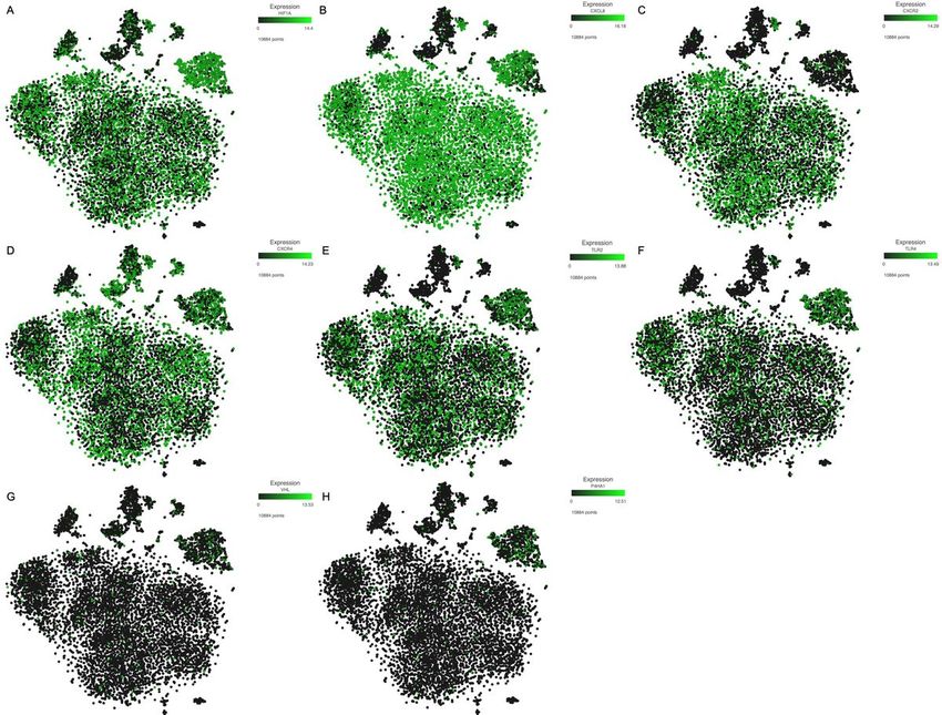
Once the blood cell populations were identified, we looked for HIF1α gene expression. As shown in Figure
2A, HIF1α gene was expressed in all myeloid lineages to a greater extent than in lymphoid cells. This was
particularly evident in the mature monocyte population. Since HIF1α is a transcription factor that acts as
a trans regulator, we searched for HIF1α-regulated genes potentially involved in COVID–19 immunity.
Among these immune related genes, we found an increased expression of CXCL8 or Interleukin–8, a
chemokine involved in the migration of mature neutrophils to the site of infection in most myeloid cell
subsets, and almost no expression in lymphoid populations and monocytoid precursors (Figure 2B). In
keeping with the increased expression of CXCL8, the genes for chemokine receptors CXCR2 (Figure 2C)
and CXCR4 (Figure 2D), and also CXCR1 were also expressed at increased levels in most myeloid
lineages. It is noteworthy that lymphoid cells did express the CXCR4 gene, but showed no expression of
the CXCR2 gene, which can explain the exacerbated inflammatory response characteristic of these
Page 6/14patients 18. Interestingly, we found expression of Toll like receptor–2 and –4 (TLR2 and TLR4) in most
myeloid populations, which could be related to SARS-CoV–2 sensing (Figure 2E and 2F).
Along with the identified genes regulated by HIF1α, we found expression of genes related to metabolism
such as solute carrier family 2 member 3 (SLC2A3) also known as GLUT3, 6-phosphofructo–2-kinase
(PFKFB3), phosphoglycerate kinase 1 (PGK1) and glyceraldehyde–3-P-dehydrogenase (GAPDH) (Figure
3). This latter related with the neutrophil survival through the inhibition of their programed cell death 19.
We also found expression of superoxide dismutase 2 (SOD2), which is involved in the metabolism of
reactive oxygen species 20, vimentin (VIM) a type III intermediate filament and plasminogen activator
urokinase receptor (PLAUR), which is related to plasminogen activation.
Considering that HIF1α function is controlled by different factors, we also evaluated the expression of the
Von Hippel Lindau (VHL) and prolyl–4-hydroxylase (P4HA1) genes, two of the main inhibitors of HIF1α
function. Indeed, these two molecules are involved in the ubiquitination and degradation of HIF1α. In
keeping with the increased expression of HIF1α and target genes observed, we found that neither VHL nor
P4HA1 were expressed by the peripheral blood cells of critically ill COVID–19 patients (Figure 2G and 2H).
We next evaluated the potential interaction between HIF1α and their transcriptional targets by assessing
the simultaneous presence of their mRNAs in the same single cells (Figure 4). As shown in Figure 4A, a
significant proportion of myeloid cells co-expressed HIF1α and CXCL8. We also observed a predominance
of cells co-expressing HIF1α and TLR2 (Figure 4B) as well as HIF1α and SOD2 (Figure 4C). It is
noteworthy that among the different myeloid subsets, mature monocytes were the ones that exhibiting
co-expression of most of the analyzed genes.
Finally, we evaluated the expression of HIF1α and its transcriptional targets in peripheral blood cells from
healthy donors. To do so, we analyzed 10X Genomics publicly available datasets. We observed that
HIF1α expression was lower as compared to the expression observed in COVID–19 patients (Figure 5).
Similar results were observed for genes such as CXCL8, CXCR2, PLAUR, TLR4 and SOD2 (Figure 5).
Discussion
Hypoxia and inflammation are unequivocally linked 21 and are two of the main physiological
consequences of SARS-CoV–2 infection, particularly in severe cases. In this study we present scRNAseq
data regarding HIF1α-related gene expression in peripheral blood leukocytes from critically ill COVID–19
and characterized the different cell subpopulations. HIF1α is a heterodimeric transcription factor sensitive
to oxygen and induced under hypoxic conditions 22. The HIF1α trans element can regulate the expression
of CXCL8 23, CXCR1, CXCR2 24 and CXCR4 25. CXCL8 expression can be stimulated by interleukin (IL) 6,
TNFα, hypoxia 26 and viral infection 27 in cells such as monocytes, neutrophils, epithelial cells and
fibroblasts 28. CXCL8 is a chemokine that exerts its pro-inflammatory functions throughout the CXCR1
and CXCR2 receptors. CXCL8 and its receptors contribute to pathogen elimination, through the transient
Page 7/14activation of ERK, AKT, SRC and FAK leading to activation of neutrophils 26. The expression of CXCL8,
which is present in COVID–19 patients, is considered a potential prognostic factor in acute respiratory
distress syndrome (ARDS) 29 and lung injury 30.
The SARS-CoV–2 viral entry depends upon binding of viral spike (S) protein to the host cell surface
protein angiotensin-converting enzyme 2 (ACE2) 20. The immunopathological outcomes are most likely
induced by the host-virus interaction. The interaction between viral antigen and host immune cells results
in an exacerbated inflammatory response 31. In the present study, we also found high expression of both
TLR2 and TLR4 genes in peripheral blood leukocytes of severe COVID–19 cases. The viral Spike protein
can be recognized by TLR2 32 and TLR431, which are up-regulated in the presence of another coronavirus
such as SARS-CoV 33. TLR4 constitutes one of the most efficient innate immune receptors, triggering pro-
inflammatory responses after binding to the pathogenic ligand, and this interaction could be useful for
developing drugs 31,34.
Metabolic reprograming of innate immune cells occurs during hyperinflammatory states. Immune cells
contribute to systemic changes in metabolism by altering their metabolic profiles in response to the
immunological state. Therefore, therapeutic modulation of immune cell metabolism could alter the
inflammatory state and thus improve patient prognosis 35. Inflammation and hypoxia are inherently
linked, and hypoxia is a well-known glycolysis driver as oxygen deficit results in limited OXPHOS 36.
Previous studies have shown that the molecular mechanisms underlying the switch from OXPHOS to
glycolysis during innate immune cell response require HIF1α 35. Among the HIF1α responsive genes, we
found expression of those related to carbohydrate metabolism, such as SLC2A3/GLUT3, PFKFB3, PGK1
and GAPDH 37. HIF signaling pathway activation in neutrophils results in an increased survival of these
cells, β2 integrin expression, production of antimicrobial peptides and glycolysis. Neutrophils use high
rates of Warburg-like glycolysis for ATP generation. The Absence of HIF1α leads to reduced ATP pools
resulting in a profound impairment of the inflammatory response 38. HIF1α can also regulate nitric oxide
production, pentose phosphate pathway, OXPHOS and arginase metabolism 35. Overall,
immunometabolism is now considered an indispensable regulator of immunity, with HIF1α playing a
central role, modulating the function of various immune cell subsets 38. The expression of HIF1α has
been previously found to be a sepsis marker 35.
HIF1α participates in the regulation of a plethora of cellular events such as metabolism of ROS trough the
regulation of SOD2 39, the regulation of cytoskeleton trough VIM type III filament, which also participates
in inflammation 40, and PLAUR which activates plasminogen and activates a cascade of extracellular
proteases 41. Interestingly, the expression of this gene could be used as a predictor of severe respiratory
failure 42 which is consistent with our results.
In conclusion, in the present study, we have demonstrated the expression of HIF1α and its
transcriptionally regulated genes, in myeloid cells, including both mature and immature subsets, present
in peripheral blood of critically ill COVID–19 patients. The up-regulation and participation of HIF1α in
Page 8/14relevant events such as inflammation, immunometabolism, and TLR supports its use as molecular
marker for COVID–19 severity and as a potential candidate for targeted therapy.
Declarations
Acknowledgments
DMR is a recipient of the National Council for Science and Technology Fellowship “Catedra CONACyT”
program. This work was partially supported by grants 289499 from Fondos Sectoriales Consejo Nacional
de Ciencia y Tecnologia Mexico and R-2015-785-015 from Instituto Mexicano del Seguro Social (MM). We
would like to thank to Xiaowen Wang from Partek Inc. for the exceptional technical support provided.
Author contributions
MM, DMR and KTP conceived, designed and coordinated the project, performed scRNAseq experiments,
analyzed, discussed data and prepared the manuscript. EV, HM, CRGB, JT, AM, NWR, FBF, AFH, CRR, EPM,
GSR, SVP, CML, RCG, LBA, RS, PPS and ACG performed scRNAseq experiments, analyzed, discussed and
interpreted biological data, wrote the paper.
CLM, JCG, EFO, GFP, provided the biological sample, retrieved the immune cells and collect clinical data.
Competing interest
RCG and CML work for Analitek S.A. de C.V. which supplied research reagents. The rest of the authors
declare not competing interests.
References
1. Bi Q, Wu Y, Mei S, et al. Epidemiology and transmission of COVID-19 in 391 cases and 1286 of their
close contacts in Shenzhen, China: a retrospective cohort study. Lancet Infect Dis. 2020.
2. Coronavirus disease (COVID-2019) situation reports 176. 2020.
3. Jose RJ, Manuel A. COVID-19 cytokine storm: the interplay between inflammation and coagulation.
Lancet Respir Med. 2020;8(6):e46-e47.
4. Sarkar M, Niranjan N, Banyal PK. Mechanisms of hypoxemia. Lung India. 2017;34(1):47-60.
5. Kashani KB. Hypoxia in COVID-19: Sign of Severity or Cause for Poor Outcomes. Mayo Clin Proc.
2020;95(6):1094-1096.
6. Xie J, Covassin N, Fan Z, et al. Association Between Hypoxemia and Mortality in Patients With
COVID-19. Mayo Clin Proc. 2020;95(6):1138-1147.
7. Cavezzi A, Troiani E, Corrao S. COVID-19: hemoglobin, iron, and hypoxia beyond inflammation. A
narrative review. Clin Pract. 2020;10(2):1271.
Page 9/148. Ottestad W, Seim M, Maehlen JO. COVID-19 with silent hypoxemia. Tidsskr Nor Laegeforen.
2020;140(7).
9. Choudhury R. Hypoxia and hyperbaric oxygen therapy: a review. Int J Gen Med. 2018;11:431-442.
10. Majmundar AJ, Wong WJ, Simon MC. Hypoxia-inducible factors and the response to hypoxic stress.
Mol Cell. 2010;40(2):294-309.
11. Hoogendijk AJ, Pourfarzad F, Aarts CEM, et al. Dynamic Transcriptome-Proteome Correlation
Networks Reveal Human Myeloid Differentiation and Neutrophil-Specific Programming. Cell Rep.
2019;29(8):2505-2519 e2504.
12. Grassi L, Pourfarzad F, Ullrich S, et al. Dynamics of Transcription Regulation in Human Bone Marrow
Myeloid Differentiation to Mature Blood Neutrophils. Cell Rep. 2018;24(10):2784-2794.
13. Novershtern N, Subramanian A, Lawton LN, et al. Densely interconnected transcriptional circuits
control cell states in human hematopoiesis. Cell. 2011;144(2):296-309.
14. Lambert C, Preijers F, Yanikkaya Demirel G, Sack U. Monocytes and macrophages in flow: an ESCCA
initiative on advanced analyses of monocyte lineage using flow cytometry. Cytometry B Clin Cytom.
2017;92(3):180-188.
15. Itelman E, Wasserstrum Y, Segev A, et al. Clinical Characterization of 162 COVID-19 patients in Israel:
Preliminary Report from a Large Tertiary Center. Isr Med Assoc J. 2020;22(5):271-274.
16. Tang-Huau TL, Gueguen P, Goudot C, et al. Human in vivo-generated monocyte-derived dendritic cells
and macrophages cross-present antigens through a vacuolar pathway. Nat Commun.
2018;9(1):2570.
17. Taniguchi-Ponciano K, Vadillo E, Lopez-Macias C, et al. A shift towards an immature myeloid profile
in peripheral blood of critically ill COVID-19 patients. Research Square. 2020:1-20.
18. Sadiku P, Walmsley SR. Hypoxia and the regulation of myeloid cell metabolic imprinting:
consequences for the inflammatory response. EMBO Rep. 2019;20(5).
19. Walmsley SR, Print C, Farahi N, et al. Hypoxia-induced neutrophil survival is mediated by HIF-1alpha-
dependent NF-kappaB activity. J Exp Med. 2005;201(1):105-115.
20. Hoffmann M, Kleine-Weber H, Schroeder S, et al. SARS-CoV-2 Cell Entry Depends on ACE2 and
TMPRSS2 and Is Blocked by a Clinically Proven Protease Inhibitor. Cell. 2020;181(2):271-280 e278.
21. Vanderhaeghen T, Vandewalle J, Libert C. Hypoxia-inducible factors in metabolic reprogramming
during sepsis. FEBS J. 2020;287(8):1478-1495.
22. Masoud GN, Li W. HIF-1alpha pathway: role, regulation and intervention for cancer therapy. Acta
Pharm Sin B. 2015;5(5):378-389.
23. Imtiyaz HZ, Simon MC. Hypoxia-inducible factors as essential regulators of inflammation. Curr Top
Microbiol Immunol. 2010;345:105-120.
24. Maxwell PJ, Gallagher R, Seaton A, et al. HIF-1 and NF-kappaB-mediated upregulation of CXCR1 and
CXCR2 expression promotes cell survival in hypoxic prostate cancer cells. Oncogene.
2007;26(52):7333-7345.
Page 10/1425. Ishikawa T, Nakashiro K, Klosek SK, et al. Hypoxia enhances CXCR4 expression by activating HIF-1 in
oral squamous cell carcinoma. Oncol Rep. 2009;21(3):707-712.
26. Ha H, Debnath B, Neamati N. Role of the CXCL8-CXCR1/2 Axis in Cancer and Inflammatory Diseases.
Theranostics. 2017;7(6):1543-1588.
27. Tang FS, Van Ly D, Spann K, et al. Differential neutrophil activation in viral infections: Enhanced TLR-
7/8-mediated CXCL8 release in asthma. Respirology. 2016;21(1):172-179.
28. Qazi BS, Tang K, Qazi A. Recent advances in underlying pathologies provide insight into interleukin-8
expression-mediated inflammation and angiogenesis. Int J Inflam. 2011;2011:908468.
29. Coperchini F, Chiovato L, Croce L, Magri F, Rotondi M. The cytokine storm in COVID-19: An overview
of the involvement of the chemokine/chemokine-receptor system. Cytokine Growth Factor Rev.
2020;53:25-32.
30. Russo RC, Garcia CC, Teixeira MM, Amaral FA. The CXCL8/IL-8 chemokine family and its receptors in
inflammatory diseases. Expert Rev Clin Immunol. 2014;10(5):593-619.
31. Choudhury A, Mukherjee S. In silico studies on the comparative characterization of the interactions
of SARS-CoV-2 spike glycoprotein with ACE-2 receptor homologs and human TLRs. J Med Virol.
2020.
32. Dosch SF, Mahajan SD, Collins AR. SARS coronavirus spike protein-induced innate immune response
occurs via activation of the NF-kappaB pathway in human monocyte macrophages in vitro. Virus
Res. 2009;142(1-2):19-27.
33. Hu W, Yen YT, Singh S, Kao CL, Wu-Hsieh BA. SARS-CoV regulates immune function-related gene
expression in human monocytic cells. Viral Immunol. 2012;25(4):277-288.
34. Choudhury A, Mukherjee S. In silico studies on the comparative characterization of the interactions
of SARS-CoV-2 spike glycoprotein with ACE-2 receptor homologs and human TLRs. Journal of
medical virology. 2020:10.1002/jmv.25987.
35. Fitzpatrick SF. Immunometabolism and Sepsis: A Role for HIF? Front Mol Biosci. 2019;6:85.
36. Corcoran SE, O'Neill LA. HIF1alpha and metabolic reprogramming in inflammation. J Clin Invest.
2016;126(10):3699-3707.
37. Liu W, Shen SM, Zhao XY, Chen GQ. Targeted genes and interacting proteins of hypoxia inducible
factor-1. Int J Biochem Mol Biol. 2012;3(2):165-178.
38. Krzywinska E, Stockmann C. Hypoxia, Metabolism and Immune Cell Function. Biomedicines.
2018;6(2).
39. Gao YH, Li CX, Shen SM, et al. Hypoxia-inducible factor 1alpha mediates the down-regulation of
superoxide dismutase 2 in von Hippel-Lindau deficient renal clear cell carcinoma. Biochem Biophys
Res Commun. 2013;435(1):46-51.
40. Su L, Pan P, Yan P, et al. Role of vimentin in modulating immune cell apoptosis and inflammatory
responses in sepsis. Sci Rep. 2019;9(1):5747.
Page 11/1441. Gilder AS, Natali L, Van Dyk DM, et al. The Urokinase Receptor Induces a Mesenchymal Gene
Expression Signature in Glioblastoma Cells and Promotes Tumor Cell Survival in Neurospheres. Sci
Rep. 2018;8(1):2982.
42. Rovina N, Akinosoglou K, Eugen-Olsen J, Hayek S, Reiser J, Giamarellos-Bourboulis EJ. Soluble
urokinase plasminogen activator receptor (suPAR) as an early predictor of severe respiratory failure
in patients with COVID-19 pneumonia. Crit Care. 2020;24(1):187.
Figures
Figure 1
t-distributed stochastic neighbor embedding map (t-SNE) showing the identification of 12 cell clusters in
critically ill COVID-19 patients.
Page 12/14Figure 2
Expression levels of HIF1α and transcriptionally regulated target genes in peripheral blood cell lineages.
Figure 3
Violin plots from HIF1α transcriptionally regulated genes.
Page 13/14Figure 4
Simultaneous expression of HIF1α and target genes on the same single cell.
Figure 5
Cell populations and HIF1α gene expression identified in peripheral blood cells from healthy individuals.
Page 14/14También puede leer