Statement Brazilian Position Statement on the Use Of Multimodality Imaging in Cardio-Oncology 2021
←
→
Transcripción del contenido de la página
Si su navegador no muestra la página correctamente, lea el contenido de la página a continuación

Melo et al.
         Brazilian Position Statement on the Use Of Multimodality Imaging in Cardio-Oncology - 2021

         Statement

    Brazilian Position Statement on the Use Of Multimodality
    Imaging in Cardio-Oncology – 2021

    Development: Cardiovascular Imaging Department (Departamento de Imagem Cardiovascular – DIC) of the
    Brazilian Society of Cardiology (Sociedade Brasileira de Cardiologia – SBC)
    Norms and Guidelines Council (2020-2021): Brivaldo Markman Filho, Antonio Carlos Sobral Sousa, Aurora
    Felice Castro Issa, Bruno Ramos Nascimento, Harry Correa Filho, Marcelo Luiz Campos Vieira
    Norms and Guidelines Coordinator (2020-2021): Brivaldo Markman Filho

    Statement Authors: Marcelo Dantas Tavares de Melo,1 Marcelo Goulart Paiva,2 Maria Verônica Câmara
    Santos,3 Carlos Eduardo Rochitte,4,5 Valéria de Melo Moreira,5 Mohamed Hassan Saleh,4,6 Simone Cristina
    Soares Brandão,7,8 Claudia Cosentino Gallafrio,9 Daniel Goldwasser,10,11,12 Eliza de Almeida Gripp,13
    Rafael Bonafim Piveta,14 Tonnison Oliveira Silva,15 Thais Harada Campos Espirito Santo,16,17 Waldinai
    Pereira Ferreira,18 Vera Maria Cury Salemi,4 Sanderson A. Cauduro,19 Silvio Henrique Barberato,20,21 Heloísa
    M. Christovam Lopes,22 José Luiz Barros Pena,23 Heron Rhydan Saad Rached,24 Marcelo Haertel Miglioranza,25,26
    Aurélio Carvalho Pinheiro,27 Bárbara Athayde Linhares Martins Vrandecic,28 Cecilia Beatriz Bittencourt Viana
    Cruz,4 César Higa Nomura,4,29 Fernanda Mello Erthal Cerbino,30,31 Isabela Bispo Santos da Silva Costa,32
       Otavio Rizzi Coelho-Filho,33 Adriano Camargo de Castro Carneiro,5 Ursula Maria Moreira Costa Burgos,34
    Juliano Lara Fernandes,35,36 Marly Uellendahl,31,37 Eveline Barros Calado,38 Tiago Senra,6,29 Bruna Leal
    Assunção,32 Claudia Maria Vilas Freire,39,40 Cristiane Nunes Martins,28 Karen Saori Shiraishi Sawamura,5,14,41
    Márcio Miranda Brito,42,43 Maria Fernanda Silva Jardim,44 Renata Junqueira Moll Bernardes,45 Tereza Cristina
    Diógenes,46 Lucas de Oliveira Vieira,47,48 Claudio Tinoco Mesquita,13,49,50 Rafael Willain Lopes,5 Elry Medeiros,6
    Letícia Rigo,51 Valeska Leite Siqueira Marin,52,53 Marcelo José Santos,54 Gabriel Blacher Grossman,55,56 Priscila
    Cestari Quagliato,6 Monica Luiza de Alcantara,11,57,58 José Aldo Ribeiro Teodoro,59 Ana Cristina Lopes Albricker,60
    Fanilda Souto Barros,61 Salomon Israel do Amaral,62 Carmen Lúcia Lascasas Porto,63 Marcio Vinícius Lins
    Barros,64,65 Simone Nascimento dos Santos,66,67 Armando Luís Cantisano,68 Ana Cláudia Gomes Pereira Petisco,6
    José Eduardo Martins Barbosa,6 Orlando Carlos Glória Veloso,69 Salvador Spina,70 Ricardo Pignatelli,71,72 Ludhmilla
    Abrahão Hajjar,4,32 Roberto Kalil Filho,4,32 Marcelo Antônio Cartaxo Queiroga Lopes,73,74,75 Marcelo Luiz
    Campos Vieira,4,14 André Luiz Cerqueira Almeida76,77

    Universidade Federal da Paraíba,1 João Pessoa, PB – Brazil
    Hospital 9 de Julho, Cardiologia,2 São Paulo, São Paulo – Brazil
    Sociedade Brasileira de Oncologia Pediátrica,3 São Paulo, SP – Brazil
    Instituto do Coração (Incor) do Hospital das Clínicas da Faculdade de Medicina da Universidade de São Paulo (HCFMUSP),4 São Paulo, SP – Brazil
    Hospital do Coração (HCOR),5 São Paulo, SP – Brazil
    Instituto Dante Pazzanese de Cardiologia,6 São Paulo, SP – Brazil
    Hospital das Clínicas, Universidade Federal de Pernambuco,7 Recife, PE - Brazil
    Clínica Diagson Recife,8 PE – Brazil
    Instituto de Oncologia Pediátrica,9 São Paulo, SP – Brazil
    Hospital Federal de Ipanema,10 Rio de Janeiro, RJ – Brazil
    Rede D’Or São Luiz,11 Rio de Janeiro, RJ – Brazil
    Casa de Saúde São José,12 Rio de Janeiro, RJ – Brazil
    Hospital Pró-Cardíaco,13 Rio de Janeiro, RJ – Brazil
    Hospital Israelita Albert Einstein,14 São Paulo, SP – Brazil
    Hospital Cardio Pulmonar – Centro de Estudos em Cardiologia,15 Salvador, BA – Brazil
    Hospital Ana Nery,16 Salvador, BA – Brazil
    Diagnoson/Fleury,17 Salvador, BA – Brazil
    ACCamargoCancer Center – IMAGE,18 São Paulo, SP – Brazil
    Hospital Erasto Gaertner,19 Curitiba, PR – Brazil
    CardioEco Centro de Diagnóstico Cardiovascular,20 Curitiba, PR – Brazil
    Quanta Diagnóstico,21 Curitiba, PR – Brazil
    Hospital de Amor,22 Barretos, São Paulo, SP – Brazil
    Faculdade de Ciências Médicas de Minas Gerais,23 Belo Horizonte, MG – Brazil

     DOI: https://doi.org/10.36660/abc.20200266

1   Arq Bras Cardiol. 2021; [online].ahead print, PP.0-0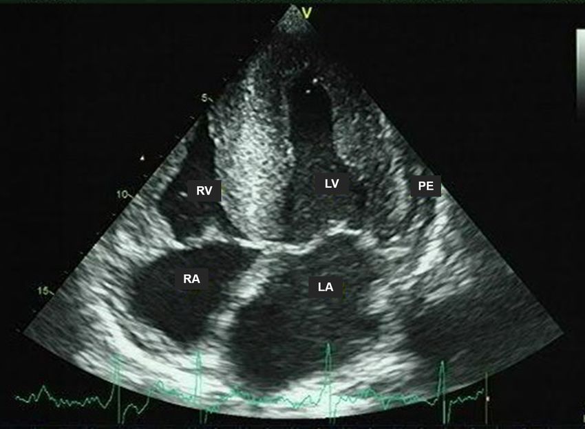
Melo et al.
                                                              Brazilian Position Statement on the Use Of Multimodality Imaging in Cardio-Oncology - 2021

                                                                                                                             Statement

Hospital Leforte Liberdade,24 São Paulo, SP – Brazil
Instituto de Cardiologia do Rio Grande do Sul – Laboratório de Pesquisa e Inovação em Imagem Cardiovascular,25 Porto Alegre, RS – Brazil
Hospital Mãe de Deus,26 Porto Alegre, RS – Brazil
Hospital Adventista de Manaus,27 Manaus, AM – Brazil
Biocor Instituto,28 Nova Lima, MG – Brazil
Hospital Sírio-Libanês,29 São Paulo, SP – Brazil
Clínica de Diagnóstico por Imagem,30 Rio de Janeiro, RJ – Brazil
Diagnósticos da América AS,31 Rio de Janeiro, RJ – Brazil
Universidade de São Paulo Instituto do Câncer do Estado de São Paulo,32 São Paulo, SP – Brazil
Universidade Estadual de Campinas (UNICAMP),33 Campinas, SP – Brazil
Universidade Tiradentes,34 Aracaju, SE – Brazil
Radiologia Clínica de Campinas,35 Campinas, SP – Brazil
Instituto de Ensino e Pesquisa José Michel Kalaf,36 Campinas, SP – Brazil
Universidade Federal de São Paulo (UNIFESP),37 São Paulo, SP – Brazil
Hospital das Clínicas da Universidade Federal de Pernambuco,38 Recife, PE – Brazil
Universidade Federal de Minas Gerais (UFMG),39 Belo Horizonte, MG – Brazil
ECOCENTER,40 Belo Horizonte, MG – Brazil
Instituto da Criança da Universidade de São Paulo (USP),41 São Paulo, SP – Brazil
Universidade Federal do Tocantins – Campus de Araguaina,42 Araguaina, TO – Brazil
Hospital Municipal de Araguaina,43 Araguaina, TO – Brazil
Hospital Samaritano de São Paulo,44 São Paulo, SP – Brazil
Instituto D’Or de Pesquisa e Ensino,45 Rio de Janeiro, RJ – Brazil
Hospital Infantil Albert Sabin,46 Fortaleza, CE – Brazil
Hospital São Rafael,47 Salvador, BA – Brazil
Rede D’Or,48 Salvador, BA – Brazil
Universidade Federal Fluminense (UFF),49 Rio de Janeiro, RJ - Brazil
Hospital Vitória,50 Rio de Janeiro, RJ – Brazil
Hospital Beneficência Portuguesa,51 São Paulo, SP – Brazil
Hospital Samaritano,52 São Paulo, SP – Brazil
Santa Casa de Misericórdia,53 São Paulo, SP – Brazil
Hospital de Câncer de Barretos,54 Barretos, SP – Brazil
Clínica Cardionuclear,55 Porto Alegre, RS – Brazil
Hospital Moinhos de Vento,56 Porto Alegre, RS – Brazil
Americas Medical City,57 Rio de Janeiro, Rio de Janeiro, RJ – Brazil
Americas Serviços Médicos,58 Rio de Janeiro, RJ – Brazil
Prenoto Medicina Diagnóstica,59 Ribeirão Preto, SP – Brazil
Centro Universitário Unihorizontes,60 Belo Horizonte, MG – Brazil
Angiolab Vitória – Diagnóstico Vascular,61 Vitória, ES – Brazil
Casa de Saúde Nossa Senhora do Carmo,62 Rio de Janeiro, RJ – Brazil
Universidade do Estado do Rio de Janeiro Faculdade de Ciências Médicas,63 Rio de Janeiro, RJ – Brazil
Mater Dei Rede de Saúde,64 Belo Horizonte, MG – Brazil
Hospital Vera Cruz,65 Belo Horizonte, MG – Brazil
Hospital Brasília – Ecocardiografia,66 Brasília, DF – Brazil
Eccos Diagnóstico Cardiovascular Avançado,67 Brasília, DF – Brazil
Hospital Barra D’Or,68 Rio de Janeiro, RJ – Brazil
Rede UHG,69 Rio de Janeiro, RJ – Brazil
Hospital Aeronáutico Central,70 Buenos Aires – Argentina
Texas Children’s Hospital, Houston,71 Texas – USA
Baylor College of Medicine, Houston,72 Texas – USA
Hospital Alberto Urquiza Wanderley – Hemodinâmica e Cardiologia Intervencionista,73 João Pessoa, PB – Brazil
Hospital Metropolitano Dom José Maria Pires,74 João Pessoa, PB – Brazil
Sociedade Brasileira de Cardiologia,75 Rio de Janeiro, RJ – Brazil
Santa Casa de Misericórdia de Feira de Santana – Cardiologia,76 Feira de Santana, BA – Brazil
Departamento de Imagem Cardiovascular da Sociedade Brasileira de Cardiologia,77 São Paulo, SP – Brazil

                                                                                                         Arq Bras Cardiol. 2021; [online].ahead print, PP.0-0   2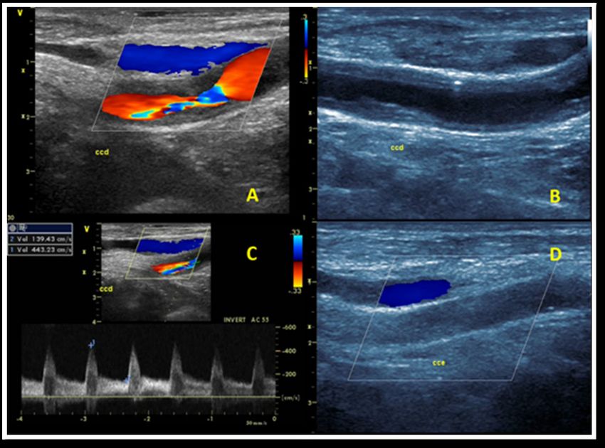
Melo et al.
         Brazilian Position Statement on the Use Of Multimodality Imaging in Cardio-Oncology - 2021

         Statement

    How to cite this Statement: Melo MDT, Paiva MG, Santos MVC, Rochitte CE, Moreira VM, Saleh MH, et
    al. Brazilian Position Statement on the Use Of Multimodality Imaging in Cardio-Oncology – 2021. Arq Bras
    Cardiol. 2021; [online]. ahead print, PP.0-0

    Note: These statements are for information purposes and should not replace the clinical judgment of a
    physician, who must ultimately determine the appropriate treatment for each patient.

    Correspondence: Sociedade Brasileira de Cardiologia – Av. Marechal Câmara, 360/330 – Centro – Rio de
    Janeiro – Postal Code: 20020-907. E-mail: diretrizes@cardiol.br

3   Arq Bras Cardiol. 2021; [online].ahead print, PP.0-0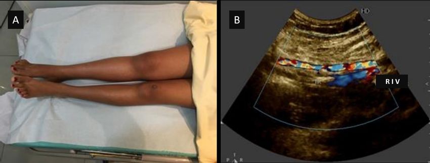
Melo et al.
Brazilian Position Statement on the Use Of Multimodality Imaging in Cardio-Oncology - 2021
Statement
Declaration of potential conflict of interests of authors/collaborators of the Brazilian Position Statement on the Use Of Multimodality Imaging in Cardio-
Oncology – 2021
If, within the last 3 years, the author/collaborator of the statement:
Participated in
clinical Was (is) a
and/or Spoke at member of Participated
Wrote
experimental events or a board of in normative Received
scientific
studies activities advisors or committees personal or Owns
Names of statement papers in
sponsored by sponsored a board of of scientific institutional stocks in
collaborators journals
pharmaceutical by industry directors of a research funding from industry
sponsored
or equipment related to this pharmaceutical sponsored by industry
by industry
companies statement or equipment industry
related to this industry
statement
Adriano Camargo de
No No No No No No No
Castro Carneiro
Ana Cláudia Gomes
No No No No No No No
Pereira Petisco
Ana Cristina Lopes
No No No No No No No
Albricker
André Luiz Cerqueira
No No No No No No No
de Almeida
Armando Luís
No No No No No No No
Cantisano
Aurélio Carvalho
No No No No No No No
Pinheiro
Bárbara Arhayde
Lihares Martins No No No No No No No
Vrandecic
Bruna Leal Assunção No No No No No No No
Carlos
No No No No No No No
Eduardo Rochitte
Carmen Lucia
No No No No No No No
Lascasas Porto
Cecilia Beatriz
No No No No No No No
Bittencourt Viana Cruz
César Higa Nomura No No No No No No No
Cláudia Cosentino
No No No No No No No
Gallafrio
Cláudia Maria Vilas
No No No No No No No
Freire
Claudio Tinoco
No No No No No No No
Mesquita
Cristiane Nunes
No No No No No No No
Martins
Daniel Goldwasser No No No No No No No
Eliza de Almeida Gripp No No No No No No No
Elry Medeiros No No No No No No No
Eveline Barros Calado No No No No No No No
Fanilda Souto Barros No No No No No No No
Fernanda Mello Erthal
No No No No No No No
Cerbino
Gabriel Blacher
No No No No No No No
Grossman
Heloísa Helena M.
No No No No No No No
Christovam Lopes
Arq Bras Cardiol. 2021; [online].ahead print, PP.0-0 4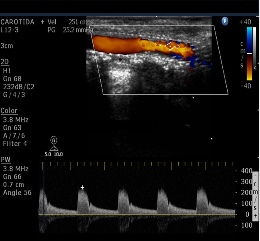
Melo et al.
         Brazilian Position Statement on the Use Of Multimodality Imaging in Cardio-Oncology - 2021

         Statement

     Heron Rhydan Saad
                                          No                  No                   No                 No   No   No   No
     Rached
     Isabela Bispo Santos
                                          No                  No                   No                 No   No   No   No
     da Silva Costa
     José Aldo Ribeiro
                                          No                  No                   No                 No   No   No   No
     Teodoro
     José Eduardo Martins
                                          No                  No                   No                 No   No   No   No
     Barbosa
     José Luiz Barros Pena                No                  No                   No                 No   No   No   No
     Juliano Lara Fernandes               No                  No                   No                 No   No   No   No
     Karen Saori Shiraishi
                                          No                  No                   No                 No   No   No   No
     Sawamura
     Letícia Rigo                         No                  No                   No                 No   No   No   No
     Lucas de Oliveira Vieira             No                  No                   No                 No   No   No   No
     Ludhmila Abrahão
                                          No                  No                   No                 No   No   No   No
     Hajjar
     Marcelo Antônio
     Cartaxo Queiroga                     No                  No                   No                 No   No   No   No
     Lopes
     Marcelo Dantas
                                          No                  No                   No                 No   No   No   No
     Tavares de Melo
     Marcelo Goulart Paiva                No                  No                   No                 No   No   No   No
     Marcelo Haertel
                                          No                  No                   No                 No   No   No   No
     Miglioranza
     Marcelo Luiz Campos
                                          No                  No                   No                 No   No   No   No
     Vieira
     Marcelo Santos                       No                  No                   No                 No   No   No   No
     Márcio Miranda Brito                 No                  No                   No                 No   No   No   No
     Márcio Vinícius Lins
                                          No                  No                   No                 No   No   No   No
     Barros
     Maria Fernanda Silva
                                          No                  No                   No                 No   No   No   No
     Jardim
     Maria Verônica Câmara
                                          No                  No                   No                 No   No   No   No
     dos Santos
     Marly Uellendahl                     No                  No                   No                 No   No   No   No
     Mohamed Hassan
                                          No                  No                   No                 No   No   No   No
     Saleh
     Mônica Luiza de
                                          No                  No                   No                 No   No   No   No
     Alcantara
     Orlando Carlos Glória
                                          No                  No                   No                 No   No   No   No
     Veloso
     Otávio Rizzi Coelho-
                                          No                  No                   No                 No   No   No   No
     Filho
     Priscila Cestari
                                          No                  No                   No                 No   No   No   No
     QuagliaA47:A68to
     Rafael Bonafim Piveta                No                  No                   No                 No   No   No   No
     Rafael Willain Lopes                 No                  No                   No                 No   No   No   No
     Renata Junqueira Moll
                                          No                  No                   No                 No   No   No   No
     Bernardes
     Ricardo Pignatelli                   No                  No                   No                 No   No   No   No
     Roberto Kalil Filho                  No                  No                   No                 No   No   No   No
     Salomon Israel do
                                          No                  No                   No                 No   No   No   No
     Amaral

5   Arq Bras Cardiol. 2021; [online].ahead print, PP.0-0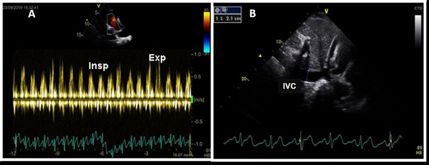
Melo et al.
                                 Brazilian Position Statement on the Use Of Multimodality Imaging in Cardio-Oncology - 2021

                                                                                                Statement

Salvador Spina         No   No      No                    No                   No                  No                 No
Sanderson A. Cauduro   No   No      No                    No                   No                  No                 No
Silvio Henrique
                       No   No      No                    No                   No                  No                 No
Barberato
Simone Cristina
                       No   No      No                    No                   No                  No                 No
Soares Brandão
Simone Nascimento
                       No   No      No                    No                   No                  No                 No
dos Santos
Tereza Cristina
                       No   No      No                    No                   No                  No                 No
Diógenes
Thais Harada Campos
                       No   No      No                    No                   No                  No                 No
Espirito Santo
Tiago Senra            No   No      No                    No                   No                  No                 No
Tonnison de Oliveira
                       No   No      No                    No                   No                  No                 No
Silva
Ursula Maria Moreira
                       No   No      No                    No                   No                  No                 No
Costa Burgos
Valéria de
                       No   No      No                    No                   No                  No                 No
Melo Moreira
Valeska Leite          No   No      No                    No                   No                  No                 No
Vera Maria Cury
                       No   No      No                    No                   No                  No                 No
Salemi
Waldinai P. Ferreira   No   No      No                    No                   No                  No                 No
Vera Maria Cury
                       No   No      No                    No                   No                  No                 No
Salemi
Waldinai P. Ferreira   No   No      No                    No                   No                  No                 No

                                                                            Arq Bras Cardiol. 2021; [online].ahead print, PP.0-0   6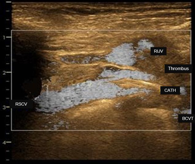
Melo et al.
          Brazilian Position Statement on the Use Of Multimodality Imaging in Cardio-Oncology - 2021

          Statement

    List of Abbreviations
     18
       F-FDG                   F-fluorodeoxyglucose
                              18
                                                                                              LVCR               Left ventricular contractile reserve
     AMI                      Acute myocardial infarction                                     LVEF               Left ventricular ejection fraction
     BMT                      Bone marrow transplantation                                     AL amyloidosis     Light-chain amyloidosis
     CHD                      Carcinoid heart disease                                         MRI                Magnetic resonance imaging
     CTX                      Cardiotoxicity                                                  SUVmax             Maximum standardized uptake value
     CVD                      Cardiovascular disease                                          NM                 Nuclear medicine
     CRT                      Catheter-related thrombosis                                     PCT                Primary cardiac tumor
     CT                       Computed tomography                                             RICAD              Radiation-induced coronary artery disease
     CAD                      Coronary artery disease                                         RIHD               Radiation-induced heart disease
     DVT                      Deep venous thrombosis                                          RIVD               Radiation-induced valve disease
     DM                       Diabetes mellitus                                               RA                 Right atrium/atrial
     ECV                      Extracellular volume                                            RV                 Right ventricle/ventricular
     GLS                      Global longitudinal strain                                      TTS                Takotsubo syndrome
     GvHD                     Graft-versus-host disease                                       99m
                                                                                                    Tc           Technetium-99m
     HF                       Heart failure                                                   TEE                Transesophageal echocardiography
     IE                       Infective endocarditis                                          TTE                Transthoracic echocardiography
     IMT                      Intima-media thickness                                          ATTR amyloidosis   Transthyretin amyloidosis
     LA                       Left atrium/atrial                                              Top2               Topoisomerase 2
     LV                       Left ventricle/ventricular                                      VTE                Venous thromboembolism

7   Arq Bras Cardiol. 2021; [online].ahead print, PP.0-0
Melo et al.
                                                                                                        Brazilian Position Statement on the Use Of Multimodality Imaging in Cardio-Oncology - 2021

                                                                                                                                                                                       Statement

Content                                                                                                             4.3.1. Coronary Artery Disease Evaluation and Follow-Up after
                                                                                                                    Radiotherapy.........................................................................................
1. General Aspects.........................................................................                         5. Cardiac Tumors and Masses.................................................
1.1. Current Situation of Cardio-Oncology in Brazil and Worldwide.........                                          5.1. Contribution from Echocardiography...............................................
1.2. Definition of Cardiotoxicity..............................................................                     5.1.1. Benign Primary Cardiac Tumors..................................................
1.3. Mechanisms of Cardiotoxicity..........................................................                         5.1.1.1. Cardiac Myxomas.....................................................................
1.4. Clinical Manifestations of Cardiotoxicity..........................................                            5.1.1.2. Papillary Fibroelastomas...........................................................
2. Myocardial Cardiotoxicity.......................................................                                 5.1.1.3. Rhabdomyomas........................................................................
2.1. Contribution from Echocardiography...............................................                              5.1.1.4. Cardiac Fibromas.....................................................................
2.1.1. Myocardial Structural and Functional Assessment of the Left                                                  5.1.1.5. Cardiac Lipomas.......................................................................
Ventricle................................................................................................           5.1.1.6. Teratomas.................................................................................
2.1.1.1. Standard Doppler Echocardiography.......................................                                   5.1.1.7. Cardiac Hemangiomas.............................................................
2.1.1.2. Myocardial Strain.....................................................................                     5.1.1.8. Cardiac Paragangliomas...........................................................
2.1.1.3. Left Ventricular Ejection Fraction by 3D Imaging.....................                                      5.1.1.9. Cardiac Schwannomas.............................................................
2.1.1.4. Contrast Echocardiography......................................................                            5.1.2. Malignant Primary Cardiac Tumors............................................
2.1.1.5. Stress Echocardiography...........................................................                         5.1.2.1. Angiosarcomas.........................................................................
2.1.1.6. Diastolic Function.....................................................................                    5.1.2.1.1. Undifferentiated Sarcomas....................................................
2.1.2. Myocardial Structural and Functional Assessment of the Right Ventricle                                       5.1.2.1.2. Rhabdomyosarcomas.............................................................
2.1.3. Late Echocardiographic Follow-up.................................................                            5.1.2.1.3. Leiomyosarcomas..................................................................
2.2. Contribution from Nuclear Medicine................................................                             5.1.2.2. Primary Cardiac Lymphomas....................................................
2.2.1. Radionuclide Ventriculography...................................................                             5.1.2.3. Primary Malignant Pericardial Mesotheliomas.........................
2.2.2. Assessment of Cardiac Sympathetic Activity with mIBG..............                                           5.1.3. Metastatic Cardiac Tumors..........................................................
2.2.3. Myocardial Metabolism – 18F-FDG PET-CT...................................                                    5.2. Contribution from Cardiac Magnetic Resonance Imaging................
2.3. Contribution from Cardiac Magnetic Resonance Imaging................                                           5.2.1. Benign Primary Tumors...............................................................
2.3.1. Assessment of Cardiotoxicity during Antineoplastic Treatment..                                               5.2.1.1. Myxomas...................................................................................
2.3.2. Cardiac Magnetic Resonance Imaging in Late Follow-up...........                                              5.2.1.2. Lipomas....................................................................................
2.3.3. Tissue Characterization by Cardiac Magnetic Resonance Imaging.                                               5.2.1.3. Papillary Fibroelastomas...........................................................
2.3.3.1. T2 Mapping...............................................................................                  5.2.1.4. Rhabdomyomas........................................................................
2.3.3.2. T1 Mapping...............................................................................                  5.2.1.5. Fibromas...................................................................................
3. Vascular Toxicity.........................................................................                       5.2.1.6. Hemangiomas...........................................................................
3.1. Contribution from Vascular Ultrasonography...................................                                  5.2.2. Malignant Tumors.......................................................................
3.1.1. Venous Thromboembolism and Cancer......................................                                      5.2.2.1. Sarcomas..................................................................................
3.1.1.1. Introduction.............................................................................                  5.2.2.2. Lymphomas..............................................................................
3.1.1.2. Epidemiology............................................................................                   5.3. Contribution from Nuclear Medicine................................................
3.1.1.3. Diagnosis of Deep Venous Thrombosis....................................                                    5.3.1. 18F-FDG PET-CT...........................................................................
3.1.1.4. Venous Ultrasound Protocols...................................................                             5.3.1.1. Cutoff SUVmax value for 18F-FDG to better differentiate benign
3.1.1.5. Differential Diagnosis of Deep Venous Thrombosis.................                                          from malignant cardiac tumors.............................................................
3.1.2. Catheter-Related Thrombosis in Patients with Cancer................                                          6. Special Situations.....................................................................
3.1.2.1. Introduction.............................................................................                  6.1. Carcinoid Heart Disease..................................................................
3.1.2.2. Risk Factors...............................................................................                6.2. Cardiac Amyloidosis........................................................................
3.1.2.3. Diagnosis and Complications...................................................                             6.2.1. Introduction................................................................................
3.1.3. Pulmonary Hypertension in Patients with Cancer......................                                         6.2.2. Clinical Types and Cardiac Involvement.....................................
4. Radiotherapy-Induced Cardiotoxicity.................................                                             6.2.3. Contribution from Echocardiography.........................................
4.1. Role of Echocardiography................................................................                       6.2.3.1. Increased Myocardial Thickness...............................................
4.1.1. Epidemiology...............................................................................                  6.2.3.2. Left Atrium................................................................................
4.1.2. Pathophysiology..........................................................................                    6.2.3.3. Diastolic Function.....................................................................
4.1.3. Initial Evaluation and Follow-Up.................................................                            6.3.3.4. Left Ventricular Systolic Function.............................................
4.1.4. Radiation-Induced Heart Disease and Role of Echocardiogram......                                             6.2.3.5. Other Findings..........................................................................
4.1.4.1. Radiation-Induced Pericardial and Myocardial Disease...........                                            6.2.3.6. Diagnostic Approach................................................................
4.1.4.2. Radiation-Induced Coronary Artery Disease............................                                      6.2.4. Contribution from Cardiac Magnetic Resonance Imaging..........
4.1.4.3. Radiation-Induced Valve Disease..............................................                              6.2.5. Contribution from Nuclear Medicine..........................................
4.2. Radiation-Induced Vascular Diseases and Role of Vascular                                                       6.3. Takotsubo Syndrome.......................................................................
Ultrasonography.....................................................................................                6.4. Siderotic Cardiomyopathy (Iron Overload)........................................
4.2.1. Radiotherapy and Atherosclerosis: Pathophysiological and Clinical                                            7. Pericardial Diseases.................................................................
Features.................................................................................................           7.1. Pericardial Tumors...........................................................................
4.2.2. Diagnosis.....................................................................................               7.1.1. Echocardiogram in Patients with Pericardial Neoplasm.............
4.2.3. Ultrasonographic Features..........................................................                          8. Cardio-Oncology in Children and Adolescents................
4.2.4. Arterial Stenosis Follow-Up.........................................................                         8.1. General Considerations...................................................................
4.3. Cardiovascular Evaluation after Radiotherapy and                                                               8.2. Main Risk Factors for the Development of Cardiotoxicity in Children and
Role of Nuclear Medicine............................................................                                Adolescents............................................................................................

                                                                                                                                                             Arq Bras Cardiol. 2021; [online].ahead print, PP.0-0             8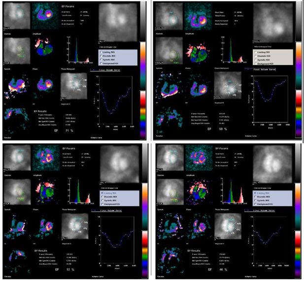
Melo et al.
           Brazilian Position Statement on the Use Of Multimodality Imaging in Cardio-Oncology - 2021

          Statement

    8.3. Cardiac Monitoring during Treatment..............................................                  Age ≥ 60 years
    8.4. Long Term Follow-up of Survivors....................................................
                                                                                                            Structural heart disease before or during treatment (ejection
    8.5. Pregnancy in Survivors of Childhood and Adolescent Cancer...........
                                                                                                         fraction: 50% to 55%, acute myocardial infarction [AMI],
    8.5.1. Cardiac Outcomes in Pregnant Survivors of Childhood and
                                                                                                         moderate/important valve disease)
    Adolescent Cancer.................................................................................
    8.5.2. Cardiovascular Monitoring Recommendation in Survivors of                                         Combination of low doses of anthracycline and trastuzumab.
    Childhood and Adolescent Cancer Wishing to Become Pregnant........
    8.6. Predisposing Situations to Thrombotic Events Related to Childhood and                           1.2. Definition of Cardiotoxicity
    Adolescent Cancer Treatment................................................................              The definition of CTX based on the degree of left ventricular
    8.6.1. Intracardiac thrombus.................................................................        ejection fraction (LVEF) reduction ignores the changes that
    8.6.2. Central Venous Catheter.............................................................
                                                                                                         precede the fall in LVEF and all other toxic effects that occur
    8.6.3. Differential Diagnosis of Intracardiac Mass................................
                                                                                                         in addition to this parameter.6-8 Lack of a more comprehensive
    8.6.3.1. Prominent Crista Terminalis.....................................................
                                                                                                         definition and, sometimes, clinical, laboratory, and imaging
    8.6.3.2. Mitral Annular Calcification.....................................................
                                                                                                         limitations to document some events in the initial stage make
    8.6.3.3. Infective Endocarditis...............................................................
                                                                                                         CTX an underdiagnosed clinical condition. The European
    8.6.3.4. Nonbacterial Thrombotic Endocarditis....................................
                                                                                                         Society of Cardiology revised in 2017 the definition of CTX
    8.6.3.5. Lambl Excrescences..................................................................
                                                                                                         to cover any structural or functional changes in the heart and
    8.6. Cardiovascular Evaluation in Case of Bone Marrow Transplantation in
                                                                                                         circulation, both in the presence or in the immediate or late
    Children and Adolescents ......................................................................
                                                                                                         post-treatment of cancer, and considered chemotherapy,
    References........................................................................................
                                                                                                         radiotherapy, or the disease itself as aggressive agents.4

                                                                                                         1.3. Mechanisms of Cardiotoxicity
    1. General Aspects                                                                                       Although we are aware of some CTX-related mechanisms,
                                                                                                         identifying the predominant mechanism remains a great
                                                                                                         challenge, as the combination of different drugs and treatment
    1.1. Current Situation of Cardio-Oncology in Brazil and
                                                                                                         protocols as well as constitutional factors inherent to each
    Worldwide
                                                                                                         patient account for a complex interaction that results in
        The estimated incidence of cancer in Brazil was 600                                              damage to the cardiovascular system (Table 1). Depending on
    thousand cases per year in 2018 and 2019.1 Only as of 2005,                                          the chemotherapy agent class, cell damage may occur directly
    the survival rate surpassed that of overall cancer mortality,                                        or indirectly and with or without potential for reversibility.9
    leading to an increased number of survivors exposed to the                                           Ewer et al.10 proposed in 2005 a classification of CTX in
    risk of cardiotoxicity (CTX), which is currently the second                                          types 1 and 2; despite being the subject of much criticism,
    leading cause of morbidity and mortality in this population.2                                        it has helped divide CTX into irreversible cell damage (type
       Cardiovascular complications resulting from cancer                                                1), attributed to anthracyclines, and reversible dysfunctions
    treatment, which are the focus of this consensus statement,                                          (type 2), attributed to trastuzumab. With the development of
    may result in premature deaths, costly hospitalizations, and                                         new anticancer therapies, including Bruton tyrosine kinase
    absence from work, leading to the need for early diagnosis                                           inhibitors, proteasome inhibitors, checkpoint inhibitors,
    and interventions.3                                                                                  among others known to be potentially cardiotoxic, it seems
       Age (children and older adults), previous myocardial or                                           that this classification proposal deserves to be revised and
    coronary heart disease, hypertension, diabetes mellitus (DM),                                        expanded.
    smoking, alcohol consumption, and sedentary lifestyle are
    factors associated with increased risk of CTX.4                                                      1.4. Clinical Manifestations of Cardiotoxicity
       Recent studies suggest that genotypic variants may modify                                            The cardiovascular clinical manifestations arising from
    the susceptibility to CTX, turning genetic mapping into a                                            cancer treatment are the tip of an iceberg whose base consists
    promising field for identification of risk subgroups.5                                               of structural and functional changes that precede signs and
       It is recommended that patients at high-risk for development                                      symptoms. For didactic purposes, we chose to divide CTX
    of CTX be considered those whose treatment includes:6                                                manifestations into three subgroups: clinical, laboratory, and
       High-dose anthracycline (doxorubicin > 250 mg/m² or                                               imaging/tracing (Table 2). It should be noted that such proposal
    epirubicin > 600 mg/m²)                                                                              may be criticized at first, as routine genetic mapping is not yet
                                                                                                         feasible to determine more accurately the culpability of the
      Radiotherapy at a dose ≥ 30 Gy (involving the heart) or
                                                                                                         phenotypic expression.
    > 2 Gy/session
                                                                                                            Anthracyclines and anti-HER2 monoclonal antibodies
         Lower doses of combined anthracycline and radiotherapy
                                                                                                         account for most documented cases of left ventricular
       Lower doses of anthracyclines or trastuzumab alone, but                                           (LV) dysfunction. Cardinale et al.11 demonstrated that the
    associated with:                                                                                     incidence of CTX for anthracycline use in a population of
       More than two cardiovascular risk factors (smoking,                                               2,625 patients was 9%, with 98% of cases occurring in the first
    hypertension, DM, dyslipidemia, obesity [during or after                                             year of treatment.11 Alkylating agents, proteasome inhibitors,
    therapy])                                                                                            and some tyrosine kinase inhibitors also cause dysfunction

9   Arq Bras Cardiol. 2021; [online].ahead print, PP.0-0
Melo et al.
                                                                           Brazilian Position Statement on the Use Of Multimodality Imaging in Cardio-Oncology - 2021

                                                                                                                                          Statement

Table 1 – Summary of the main suggested mechanisms of cardiotoxicity by group of drugs
                                  DNA double-strand break (topoisomerase IIB)
                                  Oxidative stress (reactive oxygen species)
                                  Cell membrane hyperpermeability (lipid peroxidation)
 Anthracyclines
                                  Ultrastructural changes
                                  Cytoplasmic vacuolization
                                  Apoptosis
                                  Interruption of HER-2/ERBB2 receptor signaling – Neuregulin 1
 Trastuzumab                      Inhibits cell repair
                                  Cell dysfunction
                                  Direct endothelial injury
 Cisplatin
                                  Platelet activation and aggregation
 Cyclophosphamide
                                  Coronary thrombosis
                                  Acts on the molecular signaling pathway that regulates smooth muscle tone
 5-Fluorouracil
                                  Vasospasm – vasoconstriction
                                  Inhibit nitric oxide synthase activity
 Vascular endothelial growth      Increase endothelin production
 factor
  (VEGF) inhibitors               Inhibit rho-kinase activation
                                  Vasospasm
                                  Interference with the degradation of dysfunctional proteins
 Protease inhibitors
                                  Functional changes in the myocyte
                                  Increased T-cell activity
 Immune checkpoint inhibitors
                                  Autoimmune activity in the heart muscle

Tabble – Cardiotoxicity phenotypes
                                  • Hypertension
                                  • Pulmonary hypertension
                                  • Venous and arterial embolic events
                                  • Carotid artery disease
 Clinical status                  • Heart failure/myocarditis
                                  • Pericardial effusion/pericarditis
                                  • Valve dysfunctions
                                  • Myocardial ischemia/infarction
                                  • Pericardial disease
                                  • Elevated troponins (T or I) and/or CK-MB
 Laboratory
                                  • Elevated natriuretic peptide (BNP/NT-proBNP)
                                  • Heart rhythm disorders (extrasystoles, blocks, supraventricular and ventricular tachycardias, bradyarrhythmias,
                                  increased corrected QT interval on electrocardiogram)
                                  • Dilated cardiac chambers with preserved LVEF
                                  • LVEF reduction > 10% (baseline) or > 15% (global longitudinal strain)
                                  • Left ventricular diastolic dysfunction
 Imaging/Tracing
                                  • Pericardial thickening and/or effusion
                                  • Valve dysfunctions (stenosis, failures)
                                  • Changes in imaging tests that indicate active inflammatory signs or necrosis (scintigraphy/cardiac MRI)
                                  • Changes in coronary CT angiography or calcium score that were initiated or aggravated during or after cancer treatment
                                  (chemotherapy and/or radiotherapy)
    BNP: brain natriuretic peptide; CT: computed tomography; LVEF: left ventricular ejection fraction; MRI: magnetic resonance imaging.

                                                                                                                      Arq Bras Cardiol. 2021; [online].ahead print, PP.0-0   10
Melo et al.
Brazilian Position Statement on the Use Of Multimodality Imaging in Cardio-Oncology - 2021
Statement
by means of several mechanisms.4 Severe inflammatory cardiological risk and cancer benefit. The discussed
myocarditis may be associated with immune checkpoint approaches include replacing it with lower cardiotoxic risk
inhibitors in 0.27% of patients in use of a combination of treatments, using cardioprotective measures, and even
nivolumab and ipilimumab.12 discontinuing treatment (if LVEF < 45% for anthracyclines
Coronary artery disease (CAD), clinically manifested as and < 40% for the other classes), a decision always made
stable or unstable angina or AMI, may be secondary to direct together with the oncologist.14,15
endothelial injury, acute arterial thrombosis, or vasospasm, The assessment of longitudinal systolic function,
depending on the therapeutic class that was used. Obstructive especially when advanced methods (three-dimensional [3D]
atherosclerosis, plaque rupture and coronary thrombosis, echocardiography and myocardial deformation analysis) are
annular/valvular degenerations, and pericarditis are related to unavailable, should be performed jointly. Although there are
mediastinal radiotherapy and are dependent on the radiation no reference ranges for diagnosis, a progressive decline in
dose that was used. Hypertension is closely linked to the use of the measurement of mitral annular peak systolic velocity by
endothelial growth factor inhibitors. Deep venous thrombosis tissue Doppler imaging (s’ wave) and mitral annular plane
(DVT), peripheral artery disease, and pulmonary hypertension systolic excursion (MAPSE) is significant.16
are also within the range of clinical manifestations of CTX.4 The number of times that echocardiographic imaging
is required remains controversial in the literature, varying
2. Myocardial Cardiotoxicity according to individual risk, therapeutic protocol (drugs
used and total dosage), and identification of CTX signs
and symptoms.
2.1. Contribution from Echocardiography
It is important to remember that CTX, in the form
of quantitative changes in conventional parameters for
2.1.1. Myocardial Structural and Functional Assessment assessing systolic function, may not be evident until there
of the Left Ventricle is a substantial reduction in myocardial reserve. Thus,
cardiac damage may not become apparent for years or
even decades after the end of cardiotoxic treatment, a fact
2.1.1.1. Standard Doppler Echocardiography
that is particularly applicable to adult survivors of tumors
When myocardial dysfunction was recognized as a during childhood.
potential adverse effect of cancer treatment, several strategies
were then tested to monitor myocardial function. Initially
considered to be the most accurate method, endomyocardial 2.1.1.2. Myocardial Strain
biopsy quickly fell into disuse because of its invasive nature, Strain, or deformation, is defined as the amount of
being then replaced by serial monitoring of LV systolic deformation or the fractional change in the length of a
function by noninvasive cardiovascular imaging tests. myocardial segment from initial length. Such parameter is
Echocardiography has become a consolidated method expressed as a percentage (%) and with the negative sign.17
for monitoring CTX using LVEF, as it is widely available, Two-dimensional strain imaging, deriving from speckle
cost-effective, and harmless, allowing for it to be repeated tracking, is not dependent on the angle (a limiting factor
multiple times. Additionally, it provides several other when tissue Doppler imaging is used), which makes it more
anatomical and functional findings. reproducible and more commonly used in general clinical
Administration of the Simpson method improves the practice to detect early changes in myocardial mechanics.17
estimation of ventricular volumes, overcoming the limitations Three-dimensional strain imaging represents an improvement
of fractional shortening and the Teichholz formula, obtained of the technique. In this modality, an entire pyramidal volume
from linear measurements of M-mode or two-dimensional is obtained from the apical view and then analyzed, being
(2D) echocardiography. However, sensitivity to detect small much faster than the other modalities but having lower spatial
longitudinal variations in systolic function remains low, and temporal resolution.
mainly because of frequent preload and afterload variations The fall in LVEF is a late marker of myocardial damage
during chemotherapy and intra- and inter-observer variability and translates into a poor prognosis, with reduced chance of
(one of the most accepted parameters for diagnosis of CTX), ventricular function recovery in 58% of patients, despite any
which may reach up to 10%.13 It is important to remember intervention with cardioprotective drugs. Cardiac dysfunction
that, because of those variations, tests with results outside only becomes evident when myocardial damage is significant;
the expected parameters should be repeated and confirmed therefore, absence of LVEF reduction does not exclude CTX.18,19
2 to 3 weeks after the initial finding. Thus, administration of speckle-tracking strain imaging to
The risk of CTX is considered to range from 3.6 to 11.8 analyze ventricular mechanics is gradually extending to all
times for use of cardiotoxic drugs (especially anthracyclines) heart diseases, especially those associated with the use of
if pre-treatment LVEF is between 50% and 55%. During anticancer agents, such as anthracyclines and trastuzumab.20
monitoring and after cancer treatment, CTX identification The possibility of detecting subclinical lesions has been one of
should be based on a fall > 10% in LVEF (compared to the great advantages. Overall, although early change detection
pre-treatment values) to less than 50.14 This situation is the is conceptually important, the value of actual changes must
subject of an important debate in medical teams regarding be proven to correlate with the outcomes.
11 Arq Bras Cardiol. 2021; [online].ahead print, PP.0-0Melo et al.
Brazilian Position Statement on the Use Of Multimodality Imaging in Cardio-Oncology - 2021
Statement
A review of several studies demonstrated the ability of the recommendation to repeat the tests using always the same
strain imaging to detect myocardial deformation changes device and, preferably, the same examiner.21
earlier than the fall in LVEF, either immediately after therapy An expert consensus of the American and European
infusion or in later stages.21 Cardiovascular Imaging Societies suggests that changes in
Ganame et al. 22 demonstrated the acute effects of deformation precede ventricular dysfunction.28 A reduction
anthracyclines, which are able to induce systolic dysfunction.22 > 15% in GLS immediately after or during anthracycline
The same group of investigators studied 56 patients without therapy is the most useful parameter in predicting CTX, while
any risk factors for cardiovascular disease (CVD), diagnosed a reduction > 8% will probably exclude the diagnosis of CTX
with lymphoma, leukemia, and other malignant tumors, (Figure 1).
treated only with anthracyclines (dose lower than 300 mg/ Liu et al.29 described in 2018 an algorithm to follow-up
m2), and compared them with a control group.23 After a patients treated with anticancer agents that used LVEF and GLS
mean follow-up of 5.2 years, a significant reduction in global as echocardiographic measures. In patients with LVEF > 60%,
longitudinal strain (GLS) was demonstrated at a time point the recommendation was to optimize the control of existing
when LVEF was still normal, showing that new diagnostic tools cardiovascular risk factors. Those with LVEF between 50%
are able to predict this decline early in time. and 59% and with GLS lower than -16% or at the lower limit
Sawaya et al.24 used 2D speckle tracking to demonstrate of normal were classified as preserved myocardial function;
that GLS and troponin were predictors of systolic dysfunction those with GLS greater than -16% or a 15% reduction from
in patients with breast cancer treated with anthracyclines baseline were considered to have subclinical dysfunction.
and trastuzumab. 24 Forty-three patients underwent Patients with LVEF between 40% and 49% were considered
echocardiography at baseline and at three and six months to have myocardial dysfunction; thus, this specific group was
of treatment. LVEF using the Simpson biplane method, indicated for initiation of cardioprotective therapy and a
GLS, radial and circumferential strain, and biomarkers were joint evaluation with the oncologist on the risks and benefits
assessed. In that study, GLS was able to predict CTX in seven of anticancer therapy, with an occasional dose reduction
out of nine patients, with a sensitivity of 78% and a specificity or medication change. In patients with LVEF < 40%, it is
of 79%. The event occurred at three months of follow-up in recommended that cardioprotective therapy is initiated and
one of the patients and at six months in the others. the use of a noncardiotoxic alternative therapy is discussed
Tan et al.25 examined LVEF and GLS in 19 patients with with the oncologist.
breast cancer using trastuzumab and followed-up for 34 There is no consensus on systolic function indices to
months (mean 24.7 months). They observed that changes in be monitored during treatment.14,15,29 However, recently,
ventricular function persisted for a long period, with increased the SUCCOUR (Strain sUrveillance of Chemotherapy for
LV chamber dimensions and reduced GLS throughout the improving Cardiovascular Outcomes) study was published.
entire follow-up, questioning the reversibility of the damage It showed that treatment guided by a greater than 12% drop
caused by trastuzumab.25 in the LV global longitudinal strain in patients treated with
anthracyclines is able to prevent the drop in ejection fraction
Almeida et al. evaluated 40 patients with breast cancer
and cardiotoxicity in 1 year.30 In addition to the diagnosis
who had used doxorubicin two years prior to undergoing
of CTX, the identification of GLS reduction has prognostic
an echocardiogram and compared them with 41 healthy
value, as it has been associated with higher late mortality in
women. The authors demonstrated that GLS and S’ wave of
a retrospective study involving 120 patients followed-up for
the mitral annulus were reduced in patients who underwent
21.6 ± 13.9 months.31
chemotherapy, but LVEF remained normal, suggesting the
presence of subclinical ventricular dysfunction. The authors
also showed that age and previous use of doxorubicin were 2.1.1.3. Left Ventricular Ejection Fraction by 3D Imaging
independent markers of GLS reduction.26 Three-dimensional echocardiographic imaging is the
Recently, Piveta et al.27 evaluated the role of 3D strain in method of choice for calculating LVEF during cancer
patients with breast cancer treated with anthracyclines. After treatment (Figure 2).32 By providing greater resemblance to
exposure to a low anthracycline dose (120 mg/m2), only 3D cardiac anatomy, it is much consistent with cardiac magnetic
circumferential strain and 3D area strain showed changes, resonance imaging (MRI) in the calculation of volumes, mass,
while 2D strain parameters remained unchanged.27 and LVEF.33 The 3D analysis is not dependent on geometric
A systematic review of 1,504 patients showed that a assumptions, as is the case of 2D analysis, in addition to
relative reduction of 10% to 15% in GLS from baseline was minimizing limitations related to that technique such as “apical
an important predictor for a decline in LVEF. Radial and shortening.”
circumferential strain measurements also show changes, The predominant CTX change for a consequent decrease in
but such variables are not yet routinely used. When pre- LVEF is an increase in LV end-systolic volume.15 In the oncology
chemotherapy values are not available for comparison, GLS population, studies suggest that 3D imaging is preferable to
values greater than -19% are suggestive of CTX, and the 2D imaging mainly because the former has demonstrated
association with biomarkers, especially ultrasensitive troponin, greater reproducibility and greater accuracy in the recognition
increases the sensitivity for diagnosis of CTX. It is worth noting of borderline or slightly reduced LVEF. In survivors of cancer
that reference ranges may vary depending on the software treated with anthracyclines, Armstrong et al.34 demonstrated
used in the devices and age and sex of the patients, hence that 3D imaging had greater ability to identify patients with
Arq Bras Cardiol. 2021; [online].ahead print, PP.0-0 12Melo et al.
Brazilian Position Statement on the Use Of Multimodality Imaging in Cardio-Oncology - 2021
Statement
Baseline examination (pre-chemo) LVEF < 50%
- Risk stratification and optimization and/or
- EKG ↓ GLS (< 17%)
- TTE: use 3D LVEF, if avaliable, and GLS
LVEF > 50% and normal GLS (> 17%)
Clinical evaluation with a cardiologista
Treatment optimization and clinical
follow-up
Treatment monitoring – ETT:
- Anthracyclines: baseline, at the end of treatment, 6 months after the end of
treatment, and then annuallyb
- Trastuzumab: baseline, every 3 months, and ate end of treatment
- TKI: baseline, at 3 months, and at the end of treatment
↓ LVEF > 10 points LVEF > 50% + ↓ GLS > 12% LVEF > 50% + ↓ GLS < 12%
(absolute) + LVEF < 50%c from baseline or GLS < 17%d from baseline or GLS > 17%d
Cardiotoxicity Subclinical cardiotoxicity
Evaluation with a
Do not modifiy cancer therapy + Clinical follow-up
cardiologista and repeat
cardioprotectione
TTE in 2-3 weeks for
confirmation
LVEF 40-49%
LVEF < 40%
- Abnormal systolic function:
- Abnormal systolic function:
start treatment for stage
start treatment for stage
B HFf
B HFf
- Report risks/benefits to the
- Discuss a noncartiodotoxic
oncologist
alternative for cancer
- Cancer treatment at the
treatment with the oncologist
oncologist´s discretion
Figure 1 – Clinical monitoring and management during cardiotoxic therapy.15
Adapted from: JACC Cardiovasc Imaging. 2018 Aug;11(8):1122-1131; Rev Esp Cardiol (Engl Ed) 2017; J Am Soc Echocardiogr. 2014 Sep;27(9):911-39; Arq Bras
Cardiol 2020; [online]. DOI: https:doi.org/10.36660/abc.20201006; Journal of the American College of Cardiology (2020), DOI: https://doi.org/10.1016/j.jacc.2020.11.020
a
preferably a cardio-oncologist
b
if the cumulative dose is greater than 240 mg/m2 (or equivalent), reevaluation of LVEF and SLG is recommended for each new chemotherapy cycle (50-60 mg/m2)
c
if using 3D echo, consider LVEF fall greater than 5% to less than 55% in symptomatic patients or greater than 10% in asymptomatic patients.
d
in the absence of baseline SLG (pre-chemotheraphy) for comparison, use absolute value of SLG < 17% as representative of significant change.
e
it is suggested to start cardioprotective treatment with ACE inhibitors and/or beta-blockers.
f
follow the recommendations of 2013 ACC/AHA guideline on the management of the treatment of HF stage B.
EKG: electrocardiogram; 3D: three-dimensional echocardiogram; CVRF: cardiovascular risk factores; LVEF: left ventricular ejection fraction; GLS: global longitudinal
strain; TTE: two-dimensional transthoracic echocardiogram; TKI: tyrosine kinase inhibitors; HF: heart failure; ACE: angiotensin-converting enzyme; ACC/AHA: American
College of Cardiology and American Heart Association.
LVEF < 50% than 2D imaging, with an accuracy very similar sequential assessment of LVEF over 1 year and demonstrated
to that of cardiac MRI, allowing for earlier identification of that 3D imaging had the lowest intra- and inter-observer
subclinical CTX.34 The SUCCOUR study used two criteria of temporal variability (5.6%).13 This finding suggests that, in
cardiotoxicity by preferentially 3D echocardiography: a fall of addition to being reliable, 3D imaging is a consistent and
more than 5% in patients with symptoms of heart failure, or reproducible method for evaluation of patients with cancer.32
greater than 10% in asymptomatic patients, compared with the Other papers also highlight the greater reproducibility of
baseline test for values of ejection fraction lower than 55%.30 3D imaging in the calculation of LVEF, mainly because, as
In patients undergoing chemotherapy, Thavendiranathan a semiautomatic technique for endocardial tracing, it is less
et al.13 compared different echocardiographic techniques for affected by variability in image acquisition.35
13 Arq Bras Cardiol. 2021; [online].ahead print, PP.0-0Melo et al.
Brazilian Position Statement on the Use Of Multimodality Imaging in Cardio-Oncology - 2021
Statement
Figure 2 – Example of a tree-dimensional echocardiogram with full volume analysis and estimated volumes and left ventricular ejection fraction.32
2.1.1.4. Contrast Echocardiography Civelli et al.39 prospectively measured LVCR (defined
Inadequate visualization of LV endocardial borders often as the difference between peak and rest LVEF) using low-
occurs in patients undergoing chemotherapy for breast cancer, dose dobutamine stress echocardiography during and after
particularly when following mastectomy and radiotherapy. high-dose chemotherapy in 49 women with advanced
Consequently, underestimated volumes and inaccurate breast cancer. An asymptomatic decline ≥ 5% in LVCR
LVEF determination may occur. According to international from baseline was able to predict a fall in LVEF to < 50%.39
guidelines, an ultrasound contrast agent should be used The only published systematic review on the utility of
to improve the definition of endocardial borders and the cardiac stress methods for detecting CVD in survivors of
analysis of LV function when endocardial visualization is breast cancer concluded that there seems to be evidence
limited in two or more segments. 36 Conversely, contrast that stress echocardiography is beneficial to early prognostic
agents are not recommended when LVEF is estimated on evaluation and late follow-up after anthracycline therapy.40
3D echocardiogram, as they lead to less reproducibility and Before stress echocardiography can be routinely added
greater temporal variability in LVEF compared to 3D imaging to clinical practice in cardio-oncology, further studies are
alone.13 needed to determine the best stressor, which parameters
should be measured during the test, the best time to perform
2.1.1.5. Stress Echocardiography the test according to the different types of treatment, cost-
benefit and feasibility in the oncology population, and,
Exercise or pharmacological stress echocardiography finally, the presence of incremental prognostic value over
is an established method for detecting obstructive CAD traditional parameters measured at rest (LVEF and GLS).
and subclinical changes in myocardial function. Patients
with cancer often have a decrease in global cardiovascular
reserve, attributed to the direct effects of adjuvant cancer 2.1.1.6. Diastolic Function
therapy and/or the indirect effects of lifestyle changes Abnormal parameters related to diastolic function, such
associated with treatment.37 Thus, the potential uses for as E and A waves, E/A ratio, isovolumic relaxation time,
stress echocardiography in patients undergoing cancer and myocardial performance index, have been described
therapy include the following: (a) initial investigation of early after chemotherapy. 41,42 However, longitudinal
presence of obstructive CAD in patients with intermediate- studies have not been able to reproduce the prognostic
to-high pre-test probability, noninterpretable ECG (exercise) value of those findings and there is insufficient evidence
or unable to exercise (dobutamine), especially if receiving to recommend such assessment in the diagnosis of
chemotherapy associated with ischemia or after long-term chemotherapy-induced CTX.43
radiotherapy; (b) determination of left ventricular contractile Studies have demonstrated the utility of tissue Doppler-
reserve (LVCR) as a predictor of CTX in patients with normal derived measurements in the assessment of diastolic function
rest LVEF and GLS; (c) determination of LVCR in established in patients undergoing cancer treatment. Some papers
CTX, as transient recovery of LV function during stress could have shown a reduction in tissue Doppler-derived early
indicate a better prognosis.28 Despite those potentialities, diastolic velocity (e’ wave) of the mitral annulus in patients
stress echocardiography has been scarcely used in the field treated with anthracyclines, which remained reduced during
of cardio-oncology. treatment and years later, but no predictive value for CTX
Using exercise stress echocardiography in 57 asymptomatic was demonstrated.16 Negishi et al.44 revealed that a 10%
women with normal LVEF treated for breast cancer with reduction in e’ wave velocity was observed in patients who
anthracyclines, Khouri et al.38 found a 12% reduction in stroke developed CTX after treatment with higher cumulative doses
volume and a 24% reduction in cardiac index from rest when of doxorubicin, but this parameter was not shown to have
compared to controls, suggesting impaired LVCR.38 a predictive role for LVEF fall.44
Arq Bras Cardiol. 2021; [online].ahead print, PP.0-0 14También puede leer