Programa de Doctorado en Óptica, Optometría y Visión Facultad de Óptica y Optometría - UNIVERSIDAD COMPLUTENSE DE MADRID
←
→
Transcripción del contenido de la página
Si su navegador no muestra la página correctamente, lea el contenido de la página a continuación
Programa de Doctorado en Óptica,
 Optometría y Visión
 Facultad de Óptica y Optometría
UNIVERSIDAD COMPLUTENSE DE MADRID

 1
Libro de Actas
 5º PhDay – UCM
 Facultad de Óptica y Optometría

 Madrid, 14 de octubre de 2021

© 2021 Facultad de Óptica y Optometría (UCM)
ÍNDICE
PRÓLOGO ............................................................................... 1
AGRADECIMIENTOS ............................................................. 3
COLABORADORES INSTITUCIONALES .............................. 4
EMPRESAS COLABORADORAS .......................................... 5
PONENCIA INVITADA ............................................................ 6
COMITÉ ORGANIZADOR ....................................................... 9
PROGRAMA CIENTÍFICO .................................................... 10
PONENCIAS ORALES .......................................................... 15
 Anterior and Posterior Vault Characterization and Evaluation
 throughout Time in Patients Implanted with Phakic Implantable
 Collamer Lens for Ametropia Correction. ................................... 16
 Power profile characterisation of 8 soft contact lenses for myopia
 control........................................................................................... 18
 Colorimetric quantification of free phosphate in tears of rabbits
 with induced dry eye. .................................................................... 20
 Comparative clinical evaluation of a New Isofocal intraocular
 lens against Monofocal intraocular lens. ..................................... 22
 Progressive power lenses assessment using eye tracking
 technology..................................................................................... 24
 Classification of dry eye disease with machine learning
 techniques. .................................................................................... 26
 Contact lens fitting and tear film affectation through thermal
 camera assessment. ...................................................................... 28

 1
Assessing interobserver variability in grading Meibographies
 using Pult’s scale. ......................................................................... 30
 Neuroprotective effects of melatonin in retinal ganglion cells. .... 32
 Spectral reflectance through hyperspectral imaging (HSI).......... 34
 Multifractal and statistical analysis of retinal vasculature. ......... 36
 Astigmatism tolerance in a new isofocal intraocular lens............ 38
 Effect of different dominant and non-dominant multifocal scleral
 lens combinations in vision quality. ............................................. 40
 Characterization of the meibomian glands in patients with allergic
 conjunctivitis and its relationship with histaminase. .................... 42
 Effect of the use or artificial tears on the conjunctive for different
 fixation requirements, in healthy young adults............................. 44
 BSSRDF Measurement System. .................................................... 46
 Development of near infrared reflectance scale. .......................... 48
 Effect of the elasticity module of soft contact lenses on the
 morphology and function of the meibomian glands. .................... 50
PONENCIAS EN PÓSTER .................................................... 52
 Pilot study to investigate the effect of orthokeratology contact
 lenses on rabbit eye (morphology and physiology) ...................... 53
 Development of a protocol for the characterisation of ocular
 surface microbiota in individuals with dry eye............................. 55
 Development of chronic intraocular hypertension in the rabbit:
 Effects of Intra-ocular injection of gold nanoparticles. ............... 57
 Quantitative and qualitative study of the anterior ocular pole in a
 population with multiple sclerosis. ............................................... 59
Influence of distance and hours of nearwork on accommodative
function. A 4-year longitudinal study. .......................................... 61
Impact of contact lens wear on NLRP3 gene expression:
implications for ocular frailty in middle-aged adults. .................. 63
Evaluation of retinal vasculature by OCT Angiography in type II
diabetes. ........................................................................................ 65
In vivo study of efficacy of the release of melatoninergic tear
secretagogues by contact lenses. .................................................. 67
Predictive Machine Learning applied to the CISSve Survey. ....... 69
PRÓLOGO
 Hemos llegado a la quinta edición de nuestro congreso, el 5º PhDAY-
FOO, organizado por y para los doctorandos de la Facultad de Óptica y
Optometría de la Universidad Complutense de Madrid. Se trata de un
congreso gratuito en el que estos jóvenes científicos podrán presentar sus
investigaciones al resto de sus compañeros predoctorales y a toda la
comunidad universitaria que quiera disfrutar de este evento. Apunta en tu
agenda: el 14 de octubre de 2021. En esta ocasión organizaremos un
congreso presencial, pero que permitirá acceder y participar como ponentes
a través de videoconferencia a los doctorandos con impedimentos justificados
para acudir de forma presencial. No obstante, ante la posibilidad de que la
pandemia Covid-19 pudiera impedir o dificultar su celebración presencial,
haremos los preparativos necesarios para poder pasar a formato virtual si
fuera necesario.

 A través de varias sesiones de presentaciones orales y póster, nuestros
doctorandos mostrarán la gran diversidad y riqueza de líneas de
investigación incluidas en nuestro programa de doctorado. Se
pretende difundir el trabajo desarrollado por los doctorandos de nuestra
Facultad y a la vez contribuir a mejorar sus habilidades comunicadoras como
científicos.

 Si acabas de matricularte por primera vez en el doctorado no debes
faltar a esta jornada pues es una estupenda toma de contacto con la que será
tu labor académica-investigadora en los próximos años hasta desembocar en
la presentación de tu tesis doctoral. Y si ya eres un doctorando veterano,
tu mayor experiencia te permitirá disfrutar y apreciar el gran trabajo que hay
detrás de los minutos disponibles para cada presentación.

 Además de los propios estudiantes de doctorado, serán especialmente
invitados los Estudiantes de Máster por ser ellos la cantera de futuros
doctorandos del multicisciplinar Programa de Doctorado en Óptica,
Optometría y Visión. Si eres un estudiante de Máster, en pocos meses te
encontrarás con la opción de proseguir tu trayectoria como universitario hasta
alcanzar el máximo nivel de estudios y ser Doctor, y esta jornada PhDAY-
FOO te puede ayudar a tomar esta decisión.

 1
Profesores y Personal de Administración y Servicios de la Facultad
de Óptica y Optometría seréis bienvenidos a este congreso donde podréis
conocer de la mano de sus protagonistas la interesante investigación
vinculada a nuestro Programa de Doctorado.

 Desde el Equipo Decanal de la Facultad de Óptica y Optometría y en
particular, desde mi puesto de Vicedecana de Posgrado e Investigación y
Coordinadora del Programa de Doctorado, quiero agradecer la buena acogida
que esta iniciativa ha tenido entre los doctorandos en las diversas ediciones
y en especial, agradecer al Comité Organizador toda su dedicación, ilusión y
profesionalidad. Sin todos ellos sería imposible que congreso logre las metas
propuestas.

 Beatriz Antona Peñalba
Coordinadora del programa de doctorado en Óptica, Optometría y Visión
Vicedecana de Posgrado e Investigación
Facultad de Óptica y Optometría (UCM)
bantona@ucm.es

 2

AGRADECIMIENTOS
 El Comité Organizador quisiera agradecer tanto a la Escuela de
Doctorado como a la Facultad de Óptica y Optometría de la Universidad
Complutense de Madrid, la oportunidad de realizar parte de la organización
de la 5ª Edición del PhDAY FOO – Complutense. Permitiendo de esta forma
adquirir nuevas competencias a la hora de participar en esta clase de eventos.

 Queríamos agradecer también a todos los compañeros que han
participado tanto siendo ponentes como asistentes en esta nueva edición del
PhDAY, ya que sin su colaboración todo esto no sería posible. Siendo un
congreso hecho por y para los doctorandos. Gracias nuevamente y más en
este año que ha sido atípico.

 Agradecer también a las empresas colaboradoras por el apoyo ofrecido
para llevar a cabo las jornadas PhDAY FOO y a la Asociación de Fotografía
de la Facultad por encargarse del reportaje gráfico.

 En último lugar, agradecer el esfuerzo de todo el comité científico que ha
permitido que todos los trabajos hayan sido valorados de forma objetiva, justa
y transparente, dedicando todo su tiempo a evaluar y poder otorgar así los
merecidos premios a los pertinentes participantes.

 3

COLABORADORES INSTITUCIONALES

 4
EMPRESAS COLABORADORAS

 5
PONENCIA INVITADA
“Navegando por Europa en busca de
investigación en superficie ocular”
Por: Dr. Alberto Recchioni
 En esta charla, el Dr Alberto Recchioni (AR) hablará de su experiencia
como estudiante de doctorado y luego como investigador a través de países
 como España, Alemania y Reino Unido. Tratará algunos ejemplos de las
 actividades y los estudio llevados a cabo en estos países (sobre todo de
UK) y las posibilidades que se han generado a través de networking, trabajo
 en la Salud Publica y practicas relacionadas con los temas de superficie
 ocular y ojo seco.

 Alberto Recchioni recibió su doctorado
 con una tesis titulada "Papel de la condición del ojo
 seco en la cirugía de cataratas y cirugía refractiva"
 dentro de la Red Europea de Ojo Seco (http://eden-
 ejd.eu/) (EDEN) MSCA-ITN-2014 Marie
 Skłodowska-Curie Innovative Training – European
 Joint Doctorate entre Aston University (Reino Unido)
 y la Universidad de Valencia (España).

 Actualmente, ocupa el puesto de Research Fellow en el proyecto “Fluid-
Gels as Resorbable Protective Dressings for Ocular Surface Disease” en la
University of Birmingham; dentro del Institute of Inflammation and Ageing,
colabora en un equipo multidisciplinario formado por oftalmólogos,
optometristas, científicos biomateriales e ingenieros bioquímicos. Al mismo
tiempo, forma parte del equipo clínico multidisciplinario de la clínica
Optimising Assessment in Sjögren’s Syndrome (OASIS) en la University
Hospitals Birmingham NHS Foundation Trust (UHB).

 6
COMITÉ CIENTÍFICO

 Almudena Crooke Alvarez

 Licenciado en Farmacia y Bioquímica, y Doctora en Farmacia por la
Universidad Complutense de Madrid. Profesora Titular de Universidad de la
Facultad de Óptica y Optometría de la Universidad Complutense, donde
imparte las asignaturas “Bioquímica del ojo” del Grado en Óptica y
Optometría, y “Biomarcadores y Métodos de Diagnóstico para Patologías
Oculares” del Máster en Optometría y Visión”. Su actividad investigadora se
ha centrado en la búsqueda de biomarcadores y dianas terapéuticas de las
enfermedades del ojo asociadas a la edad.

 Jesús Carballo Álvarez

 Doctor en Óptica, Optometría y Visión por la Universidad Complutense de
Madrid (UCM); diplomado en Óptica y Optometría por la UCM y licenciado en
Psicología Clínica por la UNED. Actualmente, pertenece al Departamento de
Optometría y Visión de la Facultad de Óptica y Optometría de la UCM y al
grupo de investigación Visión Aplicada

 Su actividad docente, asistencial e investigadora en los últimos años versa
principalmente sobre la adaptación de lentes de contacto, córnea irregular,
función visual, manejo de miopía y lentes intraoculares multifocales. Es autor
de 20 artículos científicos indexados, diverso material docente y varios
artículos de libros.

 Oscar Gómez Calderón

 Licenciado en Ciencias Físicas por la UCM en 1994, y Doctor en Ciencias
Físicas por la UCM en 1999 con la tesis titulada “Dinámica espacio-temporal
en láseres. Formación de patrones”. Realizó una estancia postdoctoral en la
Universidad de Stanford durante 1999 y 2000. Profesor Titular de Universidad
de la Facultad de Óptica y Optometría de la UCM desde 2010 donde
desarrolla su labor docente e investigadora. Imparte docencia relacionada
con el área de óptica, fundamentalmente Óptica Física, y su investigación se
encuentra dentro del área de la óptica no lineal.

 7
José Antonio Gómez Pedrero

 Licenciado en Ciencias Físicas por la Universidad Complutense en 1994
y Doctor en Ciencias Físicas en 1999 con la tesis titulada "Caracterización de
lentes oftálmicas mediante la matriz de potencia dióptica local". Profesor
Titular de la Faculta de Óptica y Optometría adscrito a la Sección
Departamental de Óptica, donde imparte docencia en las asignaturas "Óptica
Oftálmica I","Óptica Oftálmica II" del Grado en Óptica y Optometría y
"Procesado de Imágenes" del Máster de Tecnologías Ópticas y de la Imagen.
Coautor del libro "Modern Ophthalmic Optics" y miembro del Grupo
Complutense de Óptica Aplicada desde 2004.

 José Luis Hernández Verdejo

 Doctor en Optometría y Visión por la UCM con la tesis titulada “Variación
de la Presión Intraocular debida a Maniobras Quirúrgicas Oculares. Un
estudio Animal”. Es Profesor Contratado Doctor del Departamento de
Optometría y Visión de la Facultad de Óptica y Optometría de la UCM. Su
trayectoria de investigación ha estado vinculada al impacto sobre la visión de
la enfermedad ocular y la cirugía oftalmológica. Su dedicación docente se
centra en baja visión para personas con discapacidad visual. Presenta un
claro perfil clínico con extensa experiencia en atención primaria. En la
actualidad realiza su investigación como miembro del grupo del “Clinical and
Experimental Eye Research Group” con referencia UCM-971009-GR96/20 y
sede en la Facultad de Óptica y Optometría de la UCM.

 8
COMITÉ ORGANIZADOR

Irene Martínez Alberquilla

Graduada en Óptica y Optometría y Máster en Optometría y Visión por la
Universidad Complutense de Madrid.

Cristina Pastrana Robles

Graduada en Óptica y Optometría y Máster en Optometría y Visión por la
Universidad Complutense de Madrid.

 9
PROGRAMA CIENTÍFICO

5ª Edición PhDAY- FACULTAD DE ÓPTICA Y
 OPTOMETRÍA 2021

 14 de octubre de 2021

 10
PROGRAMA CIENTÍFICO
 5ª Edición PhDAY- FACULTAD DE ÓPTICA Y OPTOMETRÍA 2021
15:00 – 15:30 h. Inauguración
• Vicerrector de Estudios: Dr. Victor Briones Dieste
 SALÓN
• Director de la EDUCM: Dr. Fernando Gascón Inchausti
 DE
• Decana FOO. Dra. Mª Isabel Sánchez Pérez
 ACTOS
• Vicedecana de Posgrado e Investigación y Coordinadora del Programa de
 Doctorado. Dra. Beatriz Antona Peñalba
 15:30 – 16:30 h. COMUNICACIONES PRIMERA SESIÓN. ORAL 1
SALÓN DE ACTOS AULA 16
Óptica y Visión Optometría y Visión
Coordinan: Coordinan:
Dra. Almudena Crooke Álvarez Dr. José Luis Hernández Verdejo
Dr. Óscar Gómez Calderón Dr. Jesús Carballo Álvarez
Dr. José Antonio Gómez Pedrero
 16:30-17:00 h. COMUNICACIONES SEGUNDA SESIÓN. PÓSTER
HALL DE LA FACULTAD
Óptica, optometría y visión
Coordinan:
Dr. Jesús Carballo Álvarez
Dr. José Antonio Gómez Pedrero
 17:00 – 17:30 h. Descanso-café
 17:30 – 18:30 h. COMUNICACIONES TERCERA SESIÓN. ORAL 2
SALÓN DE ACTOS AULA 16
Optometría y Visión Óptica
Coordinan: Coordinan:
Dr. José Luis Hernández Verdejo Dr. Óscar Gómez Calderón
Dra. Almudena Crooke Álvarez Dr. José Antonio Gómez Pedrero
Dr. Jesús Carballo Álvarez
18:30 – 18:50 Conferencia invitada SALÓN
“Navegando por Europa en busca de investigación en superficie ocular” DE
Dr. Alberto Recchioni ACTOS
18:50- 19:00 h Entrega de Premios y Clausura

 11
DESGLOSE DEL PROGRAMA

15:00 – 15:30 h. Inauguración PhDay Facultad Óptica y
Optometría

SALÓN DE ACTOS
Vicerrectorado de Estudios. Dr. Víctor Briones Dieste
Director de la Escuela de Doctorado UCM: Dr. Fernando Gascón Inchausti
Decana de la Facultad de Óptica y Optometría. Dra. Mª Isabel Sánchez
Pérez
Vicedecana de Posgrado e Investigación y Coord. P de Doctorado. Dra.
Beatriz Antona Peñalba

15:30 – 16:30 h. COMUNICACIONES PRIMERA SESIÓN. Presentación
ORAL 1

SALÓN DE ACTOS. Línea de investigación: Óptica y Visión.
Coordinan: Dra. Almudena Crooke Álvarez; Dr. Óscar Gómez Calderón; Dr.
José Antonio Gómez Pedrero
 • Characterization of the meibomian glands in patients with allergic
 conjunctivitis and its relationship with histaminase. Jimmy Fernando
 Reyes Domínguez (on-line)
 • Colorimetric quantification of free phosphate in tears of rabbits with
 induced dry eye. Carlos Carpena Torres
 • Astigmatism tolerance in a new isofocal intraocular lens. Lidia María
 Pérez Sanz
 • Power profile characterisation of 8 soft contact lenses for myopia
 control. Julia Bodas Romero
 • Assessing interobserver variability in grading Meibographies using
 Pult’s scale. Elena Fernández Jiménez
AULA 16. Línea de investigación: Optometría y Visión.
Coordinan: Dr. José Luis Hernández Verdejo; Dr. Jesús Carballo Álvarez
 • Anterior and Posterior Vault Characterization and Evaluation
 throughout Time in Patients Implanted with Phakic Implantable
 Collamer Lens for Ametropia Correction. Jesús Beltrán Murcia.

 12
• Comparative clinical evaluation of a New Isofocal intraocular lens
 against Monofocal intraocular lens. Carla Charbel
 • Effect of different dominant and non-dominant multifocal scleral lens
 combinations in vision quality. Ana Privado Aroco
 • Contact lens fitting and tear film affectation through thermal camera
 assessment. Elena Durán Prieto
16:30-17:00 h. COMUNICACIONES SEGUNDA SESIÓN. Presentación
PÓSTER. Hall Facultad

Coordinan: Dr. Jesús Carballo Álvarez; Dr. José Antonio Gómez Pedrero
 • Pilot study to investigate the effect of orthokeratology contact lenses
 on rabbit eye (morphology and physiology). Wael Almalki
 • Development of a protocol for the characterisation of ocular surface
 microbiota in individuals with dry eye. Raquel Calderón García
 • Development of chronic intraocular hypertension in the rabbit: Effects
 of Intra-ocular injection of gold nanoparticles. Azza Dammak
 • Quantitative and qualitative study of the anterior ocular pole in a
 population with multiple sclerosis. Jorge Donís de la Torre
 • Influence of distance and hours of nearwork on accommodative
 function. A 4-year longitudinal study. Esther Mármol Errasti
 • Impact of contact lens wear on NLRP3 gene expression: implications
 for ocular frailty in middle-aged adults. Irene Martínez Alberquilla
 • Evaluation of retinal vasculature by OCT Angiography in type II
 diabetes. Nadia Minguez Caro
 • In vivo study of efficacy of the release of melatoninergic tear
 secretagogues by contact lenses. Francisco Javier Navarro Gil
 • Predictive Machine Learning applied to the CISSve Survey. Carlos
 Pérez Garmendia

17:00 – 17:30 h. Descanso.

17:30 – 18:30 h. COMUNICACIONES TERCERA SESIÓN. Presentación
ORAL 2

SALÓN DE ACTOS. Línea de investigación: Optometría y visión.
Coordinan: Dr. José Luis Hernández Verdejo; Dra. Almudena Crooke
Álvarez; Dr. Jesús Carballo Álvarez

 13
• Effect of the use or artificial tears on the conjunctive for different
 fixation requirements, in healthy young adults. Jairo Giovanni Rojas
 Yepes (on-line)
 • Effect of the elasticity module of soft contact lenses on the
 morphology and function of the meibomian glands. Jorge Giovanni
 Vargas Velasco (on-line)
 • Neuroprotective effects of melatonin in retinal ganglion cells. Miguel
 Ángel Fernández Torres
 • Classification of dry eye disease with machine learning techniques.
 Elena Diz Arias
Aula 16. Línea de investigación: Óptica.
Coordinan: Dr. Óscar Gómez Calderón; Dr. José Antonio Gómez Pedrero
 • Progressive power lenses assessment using eye tracking
 technology. Pablo Concepción Grande
 • Spectral reflectance through hyperspectral imaging (HSI). Ángela
 Gómez Manzanares
 • BSSRDF Measurement System. Pablo Santafé Gabarda
 • Development of near infrared reflectance scale. Néstor Tejedor
 Sierra
 • Multifractal and statistical analysis of retinal vasculature. Asmae
 Igalla Elyousssfi

18:30 – 19:00 Conferencia invitada. SALÓN DE ACTOS

Dr. Alberto Recchioni

19:00 h Entrega de Premios y Clausura. SALÓN DE ACTOS

 14
PONENCIAS ORALES

 Jesús Beltrán Murcia

 Julia Bodas Romero

 Carlos Carpena Torres

 Carla Charbel

 Pablo Concepción

 Elena Diz Arias

 Elena Durán Prieto

 Elena Fernández Jiménez

 Miguel Ángel Fernández Torres

 Ángela Gómez Manzanares

 Asmae Igalla El-youssfi

 Lidia María Pérez Sanz

 Ana Privado Aroco

 Jimmy Fernando Reyes Domínguez

 Jairo Giovanni Rojas Yepes

 Pablo Santafé Gabarda

 Néstor Tejedor Sierra

 Jorge Giovanni Vargas Velasco

 15
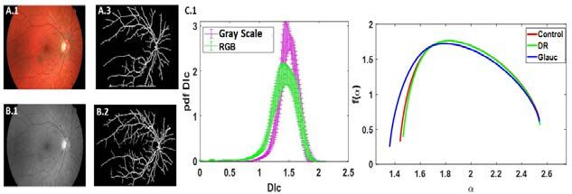
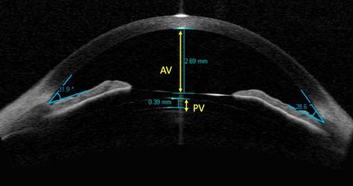
Anterior and Posterior Vault Characterization
 and Evaluation throughout Time in Patients
Implanted with Phakic Implantable Collamer
 Lens for Ametropia Correction.
 Jesús Beltrán-Murcia OD MSc 1, Vanesa Blázquez-Sánchez OD PhD 1,
 Jorge A Calvo-Sanz OD PhD2, Laureano Álvarez-Rementeria MD MSc3
 1
 Faculty of Optics and Optometry, University Complutense of Madrid,
 Spain
 2
 Staar Surgical AG 3Clínica Rementería, Madrid, Spain
 *correspondence to: jebeltra@ucm.es

Introduction: The phakic Implantable Collamer Lens- ICL pIOL (Staar
Surgical, Monrovia, CA) is a reference technique in refractive surgery for
correction of refractive errors such myopia, hyperopia or astigmatism[1]. ICL
pIOL is an epicapsular lens which will be located at the eye’s posterior
chamber behind the iris and upon the human lens. For ICL pIOL implantation
it is of great importance the fact of knowing its accurate position inside the
posterior chamber in order to learn its relationship with surrounding
structures[2] . Therefore, in this work we defined the Anterior Vault (AV) as the
distance between pIOL and corneal endothelium. Posterior Vault (PV)is also
defined as the distance between pIOL and human lens[3].

Aim: The purpose of the present study is to define AV, measure AV and PV,
to study stability of both AV and PV throughout time, get to know the possible
existing relationship among them and to evaluate security and safety of ICL
pIOL in a 12 months-follow-up once implanted.

Methods: In this retrospective study, eyes that underwent refractive surgery
implanted with ICL pIOL were analyzed. 1 month and 1 year Anterior Vault
(AV) and Posterior Vault (PV) values were measured with Visante AS-OCT
(Carl Zeiss Meditec, Inc., Ireland) postoperatively [figure 1]. All these data
obtained were processed and analyzed by using IBM SPSS Statistics V25
Software (IBM, Armonk, New York, USA).

Results: 40 eyes were analyzed. All eyes were operated by the same expert
surgeon, L.A.R.C.. 1m and 1y postop AV mean values were 2503.90±279.97
and 2560.40±278.43µm respectively with statistically significant differences
(p
56.50±134.19µm. 1m and 1y postop PV mean values were 496.63 ± 169.96
and 432.67 ± 162.74µm respectively (statistically significant differences
(p
Power profile characterisation of 8 soft
 contact lenses for myopia control.
 Julia Bodas Romero1, Laura Batres Valderas 1, Ainhoa Conde
 Rubio 1, Jesús Casas García 1, Gonzalo Carracedo
 Rodríguez 1
 1
 Deparment of Optometry and Vision, Faculty of Optics and
 Optometry, University Complutense of Madrid, Spain

 *correspondence to: jbodas@ucm.es

Introduction: Due to the myopia increases in recent years, more comfortable,
effective and safer options such as soft contact lenses for myopia control are
being investigated. Different designs such as dual focus, multifocal design or
extended depth of focus (EDOF) lenses are currently being fitted.(1, 2)
Aim: The aim of the study is to characterize the power profile of each of the 8
specific contact lenses for myopia control.
Methods: For the characterisation of the contact lenses, the NIMO TR1504
was used. The instrument is based on a technique based of quantitative
deflectometry Schlieren phase change. A reference measurement was taken
before each contact lens evaluation. The lenses were placed in the wet-
cuvette (a quartz cuvette) with saline solution, and placed in the instrument.
Twelve contact lenses of different powers of each design were measured from
-0.50 D up to -6.00 D in 0.50 D steps.
Results: Out of the 8 designs evaluated, one was a dual focus design, two
designs were EDOF, and the remaining five were multifocal designs. Optical
zone diameters between 0.93 and 3.69 mm central were obtained. It was
observed the highest myopic power and the lowest addition for the dual focus
design, while for the EDOF and multifocal designs, the additions were similar.
Conclusions: We evaluated three main soft contact lens designs specific for
myopia control (dual focus, EDOF and multifocal design). There are important
differences between designs in terms of power profile, showing different
optical zone diameters and different peripheral additions. It would be
interesting to analyze the effect of the different designs in the visual quality
and axial length elongation control.
Keywords: Soft contact lenses, myopia control, power profiles

 18
References:
[1] Anstice NS, Phillips JR. Effect of dual-focus soft contact lens wear on axial myopia progression in children.
Ophthalmology. 2011;118(6):1152-61.

[2] Bakaraju RC, Ehrmann K, Ho A. Extended depth of focus contact lenses vs. two commercial multifocals:
Part 1. Optical performance evaluation via computed through-focus retinal image quality metrics. J Optom.
2018;11(1):10-20.

 19
Colorimetric quantification of free phosphate
 in tears of rabbits with induced dry eye.
 Carlos Carpena Torres1,*, Fernando Huete Toral2, Juan
 Gonzalo Carracedo Rdríguez1
 1
 Ocupharm Research Group, Department of Optometry and
 Vision, Faculty of Optics and Optometry, Complutense
 University of Madrid, Madrid, Spain
 2
 Ocupharm Research Group, Department of Biochemistry
 and Molecular Biology, Faculty of Optics and Optometry,
 Complutense University of Madrid, Madrid, Spain
 *correspondence to: ccarpena@ucm.es

Introduction: Part of the inorganic phosphate present in the body is the
consequence of the nucleotide degradation process [1, 2]. Considering that
nucleotides such as diadenosine tetraphosphate (Ap4A), diadenosine
pentaphosphate (Ap5A), and adenosine triphosphate (ATP) manifest a higher
concentration in tears of dry eye patients, acting as molecular biomarkers [3],
it is hypothesized that free phosphate in tears could be a new biomarker easily
quantified by colorimetric methods.
Aim: To measure the free phosphate concentration in tears of rabbits with
induced dry eye compared with healthy rabbits.
Methods: An experimental, cross-sectional, and randomized study was
performed on 10 male New Zealand white rabbits. The rabbits were divided
into two groups: dry eye rabbits (n = 5) and healthy rabbits (n = 5). Dry eye
was induced by the topical instillation of benzalkonium chloride 0.2%, twice
daily, for 5 consecutive days, while the healthy rabbits received topical saline
solution as control. One hour after the last instillation, the tear samples were
collected by the Schirmer’s test for 5 min. After sample processing, the free
phosphate concentration was quantified with a commercial Malachite Green
kit by measuring the absorbance at 660 nm.
Results: The free phosphate concentration was higher in dry eye rabbits
(22.50 ± 14.06 mM) compared with healthy rabbits (18.40 ± 9.35 mM).
However, there were no statistically significant differences between both
groups (P = 0.452). Besides, there was no correlation between the free
phosphate concentration and tear volume in the total sample (r = -0.419, P =
0.066).
Conclusions: Free phosphate is proposed as a possible new molecular
biomarker to diagnose dry eye by its colorimetric detection in tears. However,

 20
future clinical studies in dry eye patients are necessary to confirm the current
findings.
Keywords: dry eye; phosphate; nucleotides.
References
[1] Fox IH. Metabolic basis for disorders of purine nucleotide degradation. Metabolism
1981;30:616-634.
[2] Yegutkin GG. Adenosine metabolism in the vascular system. Biochem Pharmacol
2021;187:114373.

[3] Carracedo G, Crooke A, Guzman-Aranguez A et al. The role of dinucleoside polyphosphates
on the ocular surface and other eye structures. Prog Retin Eye Res 2016;55:182-205.

 21
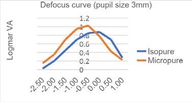
Comparative clinical evaluation of a New
 Isofocal intraocular lens against Monofocal
 intraocular lens.
 Carla Charbel1,2, Jesús Carballo Álvarez1,2, Nuria Garzón Jiménez1,2
 1
 Department of optometry and vision
 2
 Complutense University of Madrid. Faculty of Optics and Optometry
 *Correspondence to: ccharbel@ucm.es

Introduction: Cataract, lens opacification, is the common treatable cause of
loss vision and blindness worldwide.(1) Opacified lens extraction and
intraocular lens (IOL) implantation, are still the main management approach
to cataracts. In this term, monofocal IOLs are the most commonly
implanted.(2) Given the high demand from patients for spectacle
independence, multifocal IOL are becoming more and more used, providing
satisfactory far, intermediate and near vision.(2) Their concept is to create lens
that provides good vision for different distances, although for this purpose the
visual quality may decrease and photopic phenomena may appear when
compared to monofocal lenses.(2) The EDOF (Extended Depth of Focus)
technology is the most recent, and has been developed with the aim of
improving visual quality especially in intermediate vision (2) by creating a
single elongated focal point to enhance the depth of focus.(3)

Aim: The aim of the present study is to investigate visual outcomes of an
EDOF IOL (Isopure 1.2.3®) in comparison to a monofocal IOL (Micropure
1.2.3®) by the same manufacturer (PhysIOL, Belgium).

Methods: 50 cataract patients were recruited and received bilateral
implantation. They are being divided in two groups, one implanted with
Isopure 1.2.3, device under investigation and the control group implanted with
Micropure 1.2.3, based on 1:1 randomization given by the electronic data
capture. Each patient attends a total of maximum 11 visits over 12 months.
First, monocular best corrected distance visual acuity (CDVA), under photopic
luminance conditions (85 cd/m2) on the first implanted eye, will be compared
between both groups. As secondary study endpoint several parameters will
be checked as manifested refraction, monocular and binocular distance
corrected intermediate visual acuity (DCIVA), aberrometry outcomes, defocus
curve, outcomes of halos and glare simulator, binocular contrast sensitivity
under photopic and mesopic conditions (
standardized questionnaires (QoV, PRSIQ, Catquest). Statistically significant
difference was set at a level of 0.05.

Results: At the time of writing this abstract, 20 patients completed the 30-
60days visit. As predicting result, we found no statistically significant
differences for the CDVA, the halometry and the defocus curve (see figure 1),
between both groups (P-value>0.05). 9 patients completed the 75-105days
visit. As predicting result, we did not find statistically significant differences for
the defocus curve with 3 and 4.5mm pupil size. However, a better intermediate
vision with Isopure lens for both pupil size (3mm and 4.5mm) was observed.

Conclusions: The preliminary results show that the lens with isofocal design
provides better visual outcomes for intermediate distance, conserving a good
far distance quality of vision, measurements compared to a monofocal lens.

1Defocus curve comparison for pupil size 3mm.

 Keywords:
EDOF extended depth of focus, IOL intraocular lens, Isofocal.

References:
[1] Thompson J, Lakhani N. Cataracts. Prim Care - Clin Off Pract. 2015;42(3):409-23.

[2] Liu J, Dong Y, Wang Y. Efficacy and safety of extended depth of focus intraocular lenses in
cataract surgery: A systematic review and meta-Analysis. BMC Ophthalmol. 2019;19(1):1-10.

[3] Akella SS, Juthani V V. Extended depth of focus intraocular lenses for presbyopia. Current
Opinion in Ophthalmology. 2018.

 23
Progressive power lenses assessment using
 eye tracking technology.
 Pablo Concepción 1, Amelia González 1, Eva Chamorro 1 José
 Miguel Cleva 1, José Alonso 1-2 Jose Antonio Gómez-Pedrero2
 1
 Indizen Optical Technologies
 2
 Applied Optics Complutense Group, Optics Department,
 Optics and Optometry Faculty, Complutense
 University of Madrid

 *correspondence to: pconce01@ucm.es

Introduction: Presbyopia can be defined as a condition where the
accommodation of the eye is insufficient for near vision due to aging(1).
Progressive power lenses (PPL) are one of the most common solutions
nowadays to correct presbyopia. PPLs are characterized by having a smooth
and continue increase of spherical equivalent power from the upper part of the
lens to the lower portion. PPLs allows subjects to see clearly at all distances
changing the gaze position. Due to this power variation in the vertical axis,
appears unwanted astigmatism in the lateral areas of the lens(2). Unwanted
astigmatism is perceived as blur, distortion, and swim effect. The relation
between clear and blur regions of a PPL is determined by the lens design,
which is different depending on the manufacturer. Therefore, the research of
new methodologies and knowledge to evaluate the performance of PPLs are
especially important for develop new lens designs. Eye tracking technology
allows monitoring and recording eye movements using infra-red light, this
technology is used in many fields to analyze eye movements such as
marketing, driving, or evaluating reading behavior(3). The main goal of this
research is to explore the possibilities of this technology to evaluate the
performance of PPLs, specially in this work eye-tracking data has been related
with the subjective information provided by subjects to determine relationships
between characteristics of the eye movements and the PPL user’s preference.

Methods: A prospective observational double-mask study was carried out on
38 presbyopic subjects. Tobii-X3-120 (120 Hz) was used to record binocular
eye position of subjects when they were reading a text on a computer screen
with 2 types of PPL lenses with different power distributions (PPL-soft and
PPL-hard) developed ad-hoc for this research. The eye movements
parameters analyzed were fixations (number of fixations, complete fixation

 24
time, fixation duration mean) saccades (saccade duration mean and saccade
distance mean) and regressions (number of regressions). Eye movements
were analyzed for 6 different regions of the computer screen. In addition, an
analysis was carried out to find relations between characteristics of the eye
movements and the preference for a PPL.

Results: It was found a statistically significant relation between the
characteristics of the eye movements and the subjective preference for a PPL
design. Subjects that preferred the PPL-soft presented significantly worse eye
movement statistics associated with less effective eye movements than those
subjects who preferred the PPL-hard who presented significantly better eye
movements statistics associated with more effective eye movements.
Conclusions: Eye-tracking technology can be used to compare and quantify
the visual performance provided by PPL designs. Results of this research
suggest that eye tracking systems could be used as a PPL design
recommendation system through the analysis of the subjects’ eye movements
performance. This data can be applied by optometrists to determine the power
distribution of a PPL which can suited better for each subject.

Keywords: Eye-tracking, eye movements performance, progressive power
lenses
References
[1] Millodot M. Dictionary of optometry and visual science. 2009.
[2] Alonso J, Gómez-Pedrero JA, Quiroga JA. Modern Ophthalmic Optics: Cambridge University
Press; 2019.
[3] Holmqvist K, Nyström M, Andersson R, Dewhurst R, Jarodzka H, Van de Weijer J. Eye
tracking: A comprehensive guide to methods and measures: OUP Oxford; 2011.

 25
Classification of dry eye disease with
 machine learning techniques.
 Elena Diz Arias1*, Elena Fernández 2, Assumpta Peral 2,
 José A. Gómez-Pedrero 1
 1
 Applied Optics Complutense Group, Department of Optics,
 Faculty of Optics and Optometry, Complutense University of
 Madrid, Madrid, Spain.
 2
 Departament of Optometry and Vision, Faculty of Optics and
 Optometry, Complutense University of Madrid, Madrid, Spain.
 *correspondence to: elenadiz@ucm.es

Introduction: Dry eye disease (DED) is a multifactorial, chronic and
progressive disease that alters the ocular surface and the tear film. Millions of
people in the world are affected by DED. Patients suffering from this pathology
have a deteriorated quality of life and visual function. [1].

One of the most common types of DED is evaporative dry eye, this is directly
related to the function of the Meibomian Glands (MG) [2]. There are different
objective and subjective tests that allow its diagnosis and classification.
However, there is no ´Gold standard´ test or a definitive consensus among
professionals about which is the ideal set of test for its diagnosis.

In recent years, the use of artificial intelligence has represented a great
advance in the field of biomedical and health sciences. Being these promising
techniques in the prediction of diseases, either based on numerical data or
images [3].

Aim: The main objective of this study will be the prediction of the diagnosis of
dry eye disease, using objective and subjective indicators, using machine
learning models. The secondary objective will be to observe the variation of
the diagnosis by adding the parameter of the glandular contrasts of the
Meibomian glands and its possible correlation with dry eye disease.

Methods: Relevant clinical tests for the diagnosis of dry eye have been carried
out to 45 subjects (15 control, 15 contact lens wearers and 15 with Meibomian
gland pathology). Symptomatology tests, ocular surface recognition and MG
imaging were performed. The machine learning program will be trained and

 26
verified using the data obtained in these tests. In addition, a new indicator for
the diagnosis of DED will be implemented, the MG contrast.

Results: It is expected to obtain a good precision and reliability in the
diagnosis when the machine learning program is trained. It must be able
through different indicators of dry eye, to provide a specific diagnosis.
Glandular contrast can be a potential indicator for the diagnosis of DED.

Conclusions: Machine learning techniques can be advantageous when
diagnosing multifactorial pathologies such as dry eye syndrome. Meibomian
gland contrast may be a new indicator for your diagnosis.
It is intended to obtain greater precision and consensus among professionals,
which will mean an improvement in daily clinical practice.

Keywords: Dry eye disease, Meibomian glands contrast, Machine learning.

References
[1] Sullivan B.D, Crews L.A, Messmer E.M et al. Correlations between commonly used objective
signs and symptoms for the diagnosis of dry eye disease: clinical implications. Acta
ophthalmologica, 2014;92;161-166.
[2] Nichols K.K. The International Workshop on Meibomian Gland Dysfunction: Introduction.
Investigative ophthalmology & visual science. 2012;52;1917-1921.
[3] Murphy K.P.Machine learning: a probabilistic perspective. MIT press. 2012.

 27
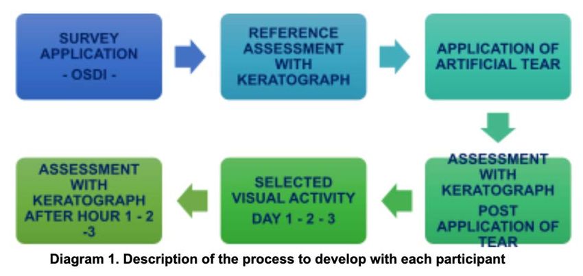
Contact lens fitting and tear film affectation
 through thermal camera assessment.
 Elena Durán-Prieto 1,2, J.M. López Alonso2,
 Jesús Carballo-Álvarez2
 1
 mark’ennovy Personalized Care
 2
 Facultad de Óptica y Optometría
 *correspondence to: eduran@ucm.es

Introduction: The contact lenses fitting is an important issue since contact
lens wear affects the tear film and could impair dry eye disease. Contact lens
design could affect dry eye due to the different thickness across them as well
as its movement on eye. For instance, contact lenses with a multifocal
design[1] tend to have variable thicknesses that are different from
conventional spherical and toric designs, which can affect comfort [2].
Aim: In this work, 4 different soft contact lens designs with different thickness
(spherical, multifocal and two toric) were analysed to evaluate and compare
the fitting through tear film stability through the thermal image of the eye,
where the lipid layer of the tear appears as dark bands, and its dynamic
evolution.
Methods: The thermal imager used is the Flir A325 model. It has been used
with a 4X close-up lens, prepared for a working distance of 7.9 mm whose
field of view is 6 ° X 4.5 °. (Figure 1). The patient places his eye about 8 mm
from the objective with his head resting on a chin rest. Contact lenses used
were made of silicone hydrogel material with low coefficient of friction
individually manufactured for each patient.

 Figure 1: From left to right: experimental set up, thermal image baseline and with
 contact lens.

 28
Videos were recorded: 1st normal blinking and break up-time tear film without
lens; 2nd with lens and 3rd, 5 and 10 minutes after removal. Videos were
analysed with MATLAB.
Results: Previous study [3] spherical and multifocal contact lenses were
analysed with thermal camera and similar records were found regardless the
optical design, but temperature decreased when contact lens was put in due
to tear film disruption. After 3 mins removal baseline temperature profile was
achieved independently of the design.
In the toric designs, preliminary results show that the thinner the contact lens
the less the tear film disruption. Dark zones, meaning less temperature are
visible in the thicker part of the toric lenses.
Moreover, the time needed to recover the baseline tear film profile increased
up to even 10 minutes in some patients, maybe due to the asymmetry of the
design (inhomogeneity of the lenses thickness), even considering the lenses
were very stable.
Conclusions: Thermal camera image is a useful tool to evaluate contact lens
fitting based on tear film stability, specially when comparing different designs.
Temperature decreases regardless lens design maybe due to tear film
disruption. In toric contact lenses designs tear film disruption seems to be
related to local thickness maybe increasing the baseline tear film profile
recovering time. It has to be taken into account that local thickness lens in
toric designs is not only affected by stability criteria but also by the
prescription. This opens new lines of research as to study interaction between
prescription and stability. Another one could be and to study the influence of
different contact lens materials.
Keywords: soft toric contact lens, tear film, thermal camera.
References
[1] Thomas G. Quinn, O. M. (9/22/2011). Review of contact lenses.
http://www.reviewofcontactlenses.com/content/c/30212/
[2] Craig, J. S. “The role of tear physiology in ocular surface temperature”. Eye, Royal College of
Ophthalmologists, Vol 14, 635-641.(2000)
[3] Durán-Prieto and J. M. López-Alonso, "Contact lens fitting through assessment of tear film
affectation with different designs by means of thermal camera," in Frontiers in Optics / Laser Science,
B. Lee, C. Mazzali, K. Corwin, and R. Jason Jones, eds., OSA Technical Digest (Optical Society of
America, 2020), paper JTu7C.4.

 29
Assessing interobserver variability in grading
 Meibographies using Pult’s scale.
 Elena Fernández Jiménez1* Elena Diz Arias2, José Antonio Gómez-Pedrero 2,
 Assumpta Peral1,
 1
 Departament of Optometry and Vision, Faculty of Optics and Optometry,
 Complutense University of Madrir, Madrid, Spain.
 2
 Applied Optics Complutense Group, Departament of Optics, Faculty of Optics and
 Optometry, Complutense University of Madrid, Madrid, Spain.
 *correspondence to: elefer06@ucm.es

Introduction: Meibomian glands (MG) are modified sebaceous glands
located in the eyelids [1], distributed along the superior and inferior tarsus and
are responsible for secreting lipids onto the tear film.
The observation of the gland morphology is key, in daily clinical practice, for
the diagnosis of associated pathologies, allowing vision professionals the
understanding, diagnosis and the subsequent treatment of these alterations.
The most used technique for the observation of MG is the non-contact
meibography with infrared (IR) light.
This imaging technique provides information of the morphologic features of
MG. Meibography allows analysis of the percentage of gland loss, in addition
to observation of morphological characteristics such as the angle of torsion,
thickness, width, length and shape of the glands [2].

In the present study, two different meibographers have been used to obtain
the MG images from a normal population. The main difference between these
devices has been the light used to visualize the glands. The first instrument
used was OCULUS Keratograph 5MTM which incorporates an IR camera to
image the Meibomian glands.
The second instrument used has been an experimental device, called Visible
Light Non-contact Meibographer (VLNCM) that uses a visible light source
combined with a red filter to obtain the MG images.
Seventeen observers, not specialized in this field, were recruited to grade the
images using the Pult and Riede Pult 5-degree scale [3].

Aim: The main objective is to assess the variability between observers when
rating the images through a standardized scale for the measurement of the
Meibomian glands.

Methods: Meibography images were taken in one random eye and in both

 30
eyelids. The superior and inferior tarsus was used to obtain the results. A total
of forty images were captured and analysed, twenty per meibographer, ten of
each eyelid. The images taken with the two devices were masked and
randomized. After this, the meibography images were shown to the seventeen
observers to be graded following the indications of the Pult and Riede-Pult
scale. A week later, the same procedure was performed, with the images
newly randomized. The seventeen observers graded the images again, in the
same way as the previous session. Therefore, the gradations were performed
twice (Session 1 and Session 2).

Results: The inter-observer variability analysis showed that there was a high
discrepancy between observers, when the images were scored using the Pult
5-degrees scale, both for the meiboscores obtained with the K5M and with the
VLNCM, for the upper and lower eyelids. This variability is greater for the
images of the lower eyelid.
Conclusions:
This variability may be due to the discrete nature of Pult’s scale, as the
difference between one grade and the next may not be discerned. Moreover,
taking into account that the predominant grades in these normal subjects are
between 0 and 2, the difference between grade 0 -1 and 1-2 is difficult to
discern, and, sometimes, the observer would give an intermediate value
between both degrees as valid.
Keywords: Meibomian glands, meibography, meibomian gland dysfunction
References:

[1] Knop E., Knop N., Millar T et al. The international workshop on meibomian gland dysfunction:
report of the subcommittee on anatomy, physiology, and pathophysiology of the meibomian
gland. Investigative ophthalmology & visual science, 2011;52; 1938-1978.
[2] Wise R. J., Sobel R. K., & Allen R. C. Meibography: A review of techniques and
technologies. Saudi Journal of Ophthalmology,2012; 26; 349-356.
[3] Pult, H., & Riede-Pult, B. H. Non-contact meibography: keep it simple but effective. Contact Lens
and Anterior Eye, 2012; 35; 77-80.

 31
Neuroprotective effects of melatonin in retinal
 ganglion cells.
 Miguel Ángel Fernández Torres1, Ana Guzman-Aranguez1
 1
 Department of Biochemistry and Molecular Biology, Faculty
 of Optics and Optometry, Complutense University, Madrid,
 Spain
 *correspondence to: miguef15@ucm.es

Introduction: Glaucoma is characterized by progressive degeneration of
retinal ganglion cells and the optic nerve, resulting in visual field loss [1].
Although the pathogenesis of glaucoma is not fully known, accumulative
evidences indicate that oxidative stress and inflammation significantly
contribute to the development and progression of this pathology. It has been
suggested that activation of NLRP3 (nucleotide binding oligomerization
domain leucine-rich repeats containing pyrin domain 3) inflammasome by
several factors, including oxidative stress, could play a key role in
inflammation and ganglion cell loss associated to glaucoma [2]. NLRP3
inflammasome is an innate immune complex. Once activated, it triggers the
cleavage and activation of caspase-1. Activated caspase-1 mediates mature
IL-1β secretion and can lead to a process of cell death called pyroptosis.
Melatonin is a neurohormone secreted by the pineal gland but it also produced
by various ocular structures such as ciliary body, retina and lens [3]. Melatonin
can be an effective antioxidant compound acting as a direct and indirect free
radical scavenger. Less information exists about its inflammatory properties
in the eye and the full range of melatonin actions in retina is still not completely
elucidated.

Aim: The purpose of this study was to analyze the potential neuroprotective
actions of melatonin on retinal ganglion cells exposed to oxidative stress. This
oxidative stress was induced by hydrogen peroxide treatment as well as by
irradiation to light emitting diode (LED) blue light. Particularly, the ability of this
neurohormone to preserve cell viability and its effect on NLRP3
inflammasome activity were evaluated.

Methods: Retinal ganglion cells R28 were exposed to hydrogen peroxide
(400 μM, 600 μM, 700 μM and 800 μM) at different times (2, 4, 6 and 24 hours)
or a blue light emitting diode (LED) light with or without melatonin
pretreatment. Cell viability under the different experimental conditions was
assessed by 3-(4,5-dimethyl-2-thiazolyl)-2,5-diphenyl-2H-tetrazolium bromide
(MTT) test. Potential cytotoxicity of melatonin administrated alone at different

 32
concentrations was also tested. Moreover, in order to analyze the NLRP3
activation, NLRP3 and caspase 1 protein levels in total cell lysates were
evaluated by western blot.

Results: Hydrogen peroxide treatment induced a remarkable time- and
concentration-dependent decrease in R28 cell viability. Similarly, exposure to
blue LED light significantly reduced cell viability. In western blot studies, higher
levels of NLRP3 and caspase 1 were found in cells exposed to oxidative stress
as compared to control (non-treated) cells indicating that oxidative stress
stimulated NLRP3 activation. R28 cells only exposed to melatonin did not
show any deleterious effect. Melatonin pre-treatment counteracted the cell
death induced by oxidative stress conditions. Likewise, melatonin prevented
NLRP3 activation since NLRP3 and caspase 1 protein levels returned to
values that resemble to those found in control cells.

Conclusions: Hydrogen peroxide and blue LED light challenge reduced R28
cell viability. Since LED light is emerging as a powerful light source, long
lasting exposure to LED light needs consideration in relation to retina
vulnerability and photochemical damage. Melatonin pre-treatment
significantly ameliorated cell damage triggered by oxidative stress and
reduced NLRP3 activation. Consequently, this neurohormone could
ameliorate pyroptosis process contributing to preserve retinal ganglion cell
survival.

Keywords: glaucoma, retina, melatonin.

References
[1] Wójcik-Gryciuk A, Skup M, Waleszczyk W. Glaucoma -state of the art and
perspectives on treatment. Restor Neurol Neurosci. 2016;34:107-23.
[2] Yerramothu P, Vijay AK, Willcox MDP. Inflammasomes, the eye and anti
inflammasome therapy. Eye (Lond). 2018; 32:491-505.
[3] Ostrin LA. Ocular and systemic melatonin and the influence of light exposure. Clinical &
experimental optometry 2019;102:99-108.

 33
Spectral reflectance through hyperspectral
 imaging (HSI).

 Ángela Gómez Manzanares1*, Antonio Álvarez Fernández-
 Balbuena 1, Daniel Vázquez Molini 1, Juan Carlos Martínez
 Antón 1, Ricardo Bernárdez Vilaboa 1, Santiago Mayorga
 Pinilla and Anto Fernández Iglesias 2
 1
 Departamento de óptica. Facultad de Óptica y Optometría.
 Universidad Complutense de Madrid.
 2
 Universidad Carlos III de Madrid.
 *correspondence to: anggomez@ucm.es

Introduction: Considering the importance of visual analysis when evaluating
the condition of a surface, recently, multispectral and hyperspectral images
(HSI) are gaining great importance in various fields of research. Some works
of art show their deterioration from changes in the pigments that the
components, so it is possible through hyperspectral analysis to determine the
state of conservation in which an artwork is, or to detect flaws that they are
imperceptible to the naked eye.

Aim: A hyperspectral image capture system has been validated obtaining the
spectral reflectance parameter of Dalí's artwork: “Two figures” with a precision
of the pixel size of the sensor used.

Methods: From the hyperspectral image capture it is possible to obtain the
reflectance parameter of a surface. For this, equation (1) applied to each
wavelength is used to later form a hyperspectral image cube.

 ( , )− ( , )
 ( , ) = ℎ ( , ) , (1)
 ℎ ( , )− ( , )

where, is the reflectance of the sample at each pixel x and y; is the
multispectral or hyperspectral image of the sample; ℎ is the multispectral
or hyperspectral image of the white material with the size of the artwork,
needed to perform the reflectance calculations. is the reflectance value

 34
También puede leer