RENAN DAL FABBRO Efeito do consumo de vinho tinto no desenvolvimento da periodontite apical induzida em ratos - Araçatuba 2021
←
→
Transcripción del contenido de la página
Si su navegador no muestra la página correctamente, lea el contenido de la página a continuación
Campus de Araçatuba

                 RENAN DAL FABBRO

Efeito do consumo de vinho tinto no desenvolvimento da
          periodontite apical induzida em ratos

                       Araçatuba

                         2021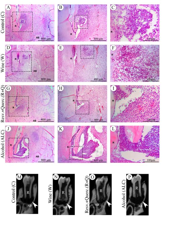
RENAN DAL FABBRO

Efeito do consumo de vinho tinto no desenvolvimento da
          periodontite apical induzida em ratos

                          Tese apresentada à Faculdade de
                          Odontologia de Araçatuba da Universidade
                          Estadual Paulista “Júlio de Mesquita Filho”
                          UNESP como parte dos requisitos para
                          obtenção do título de Doutor em
                          Endodontia.

                          Orientador: Prof. Tit. João Eduardo Gomes
                          Filho

                       Araçatuba

                         2021
Catalogação na Publicação (CIP)
Diretoria Técnica de Biblioteca e Documentação – FOA / UNESP
Fabbro, Renan Dal.
F113e Efeito do consumo de vinho tinto no desenvolvimento da
periodontite apical induzida em ratos / Renan Dal Fabbro. -
Araçatuba, 2021
55 f. : il. ; tab.
Tese (Doutorado) – Universidade Estadual Paulista,
Faculdade de Odontologia de Araçatuba
Orientador: Prof. João Eduardo Gomes Filho
1. Periodontite periapical 2. Quercetina 3. Resveratrol
4. Vinho 5. Polifenóis I. T.
Black D24
CDD 617.67
Claudio Hideo Matsumoto – CRB-8/5550Dados Curriculares
Renan Dal Fabbro
Nascimento 24/01/1992 - Capivari – São Paulo
Filiação Reinaldo Dal Fabbro
Rosa Cristina Lorenzon Dal Fabbro
2010-2014 Graduação em Odontologia
Faculdade de Odontologia de Araçatuba - Universidade
Estadual Paulista “Júlio de Mesquita Filho” (UNESP)
2015-2017 Pós-graduação stricto sensu em Ciência Odontológica
(Mestrado), área de concentração em Endodontia
Faculdade de Odontologia de Araçatuba - Universidade
Estadual Paulista “Júlio de Mesquita Filho” (UNESP)
2016-2017 Pós-graduação lato sensu em Endodontia (Especialização)
Faculdade de Odontologia de Araçatuba - Universidade
Estadual Paulista “Júlio de Mesquita Filho” (UNESP)
2017-2021 Pós-graduação stricto sensu em Ciência Odontológica
(Doutorado), área de concentração em Endodontia
Faculdade de Odontologia de Araçatuba - Universidade
Estadual Paulista “Júlio de Mesquita Filho” (UNESP)Dedicatória
Aos meus pais,
                        Reinaldo e Rosa Cristina,

      Pelo amor incondicional, força, dedicação, e pela imensa contribuição para que

eu pudesse realizar meus sonhos. Por sempre me apoiarem em todas as decisões que

tomo em minha vida. Mais do que a educação formal que vocês me ofereceram e que

sempre se esforçaram para que fosse a melhor, a formação humana foi o que de mais

importante vocês fizeram por mim. Eu só posso retribuir tentando ser o melhor filho

que pais como vocês merecem ter. Obrigado pelo maior presente que vocês puderam

me dar, a vida!

À minha irmã,
                                   Rafaela,

      Que mesmo fisicamente distante sempre esteve presente na minha formação,

torcendo e vibrando com as minhas conquistas. Tivemos tantas brigas quando crianças

que certamente ajudaram intensificar nossa amizade depois de adultos. Minha vida

não seria completa sem a sua presença!
Aos meus avós,
Cecília, Dante (in memorian), Leonice (in memorian) e
Marino (in memorian)
Fazer parte da família que vocês construíram com tanto empenho é algo
indescritível. Embora três de vocês não estejam mais presentes entre nós, sei que
continuam de olho em mim aí de cima. De vocês guardo as melhores memórias, os
melhores ensinamentos e recordo como foi importante ter vocês em minha vida. Vocês
foram uma verdadeira lição de sabedoria para mim e terei sempre um grande orgulho
de levar vossos DNA adiante.
À minha esposa,
Letícia,
Que no momento desta defesa já será minha esposa, a pessoa que compartilho
todos os detalhes da minha vida desde novembro de 2012. Esteve sempre ao meu
lado nos momentos fáceis e difíceis da vida. Agradeço do fundo do meu coração todos
os dias pelo privilégio de ter você ao meu lado. Você é uma companheira carinhosa,
engraçada, linda e leal. Seu abraço me dá o conforto que preciso para me sentir mais
tranquilo. Tenho muito orgulho do que você tem conquistado e prometo que lutarei
intensamente pela nossa felicidade. Nossa história está apenas começando, tenho
certeza de que ficaremos juntos para sempre!Agradecimentos
À Universidade Estadual Paulista “Júlio de Mesquita Filho”, na pessoa do diretor
da Faculdade de Odontologia de Araçatuba Prof. Titular Glauco Issamu Miyahara e
do vice-diretor Prof. Titular Alberto Carlos Botazzo Delbem.
Ao programa de Pós-Graduação em Ciência Odontológica da Faculdade de
Odontologia de Araçatuba – UNESP representado pelo seu coordenador Prof. Adj.
Luciano Tavares Ângelo Cintra, pela competência e qualidade na condução do
programa de pós-graduação.
À Fundação de Amparo a Pesquisa do Estado de São Paulo (FAPESP), por ter
concedido a minha bolsa de doutorado (Processo nº2017/27219-3) e por ter
fomentado meu maior sonho, que era o de fazer pesquisa no exterior, por meio da
bolsa estágio de pesquisa no exterior (Processo nº2019/05148-2).
Ao meu orientador Professor Titular João Eduardo Gomes Filho, por ter me
aceitado como aluno de Iniciação Científica lá em meados de 2012 e aberto as portas
da pesquisa científica naquele momento. Por sempre ter me apoiado e me incentivado
nos momentos difíceis, reconsiderando as solicitações à FAPESP inúmeras vezes, por
toda a paciência e ajuda que sempre teve comigo e demais orientados. Sou
eternamente grato pelos ensinamentos, pelas oportunidades e conhecimentos
repassados durante todo esse tempo. Espero um dia poder retribuir tudo.
Ao Prof. Adj. Dr. Luciano Tavares Ângelo Cintra, pela generosidade em
compartilhar seus ensinamentos durante toda a graduação e pós-graduação. Por terme aceitado no início do doutorado como seu orientado por um breve período. Pela
sua dedicação e excelente didática com as aulas. Pelo carinho com que comanda a
pós-graduação e sua pronta disposição em nos ajudar com as correções de artigos
científicos.
Ao Prof. Assist. Dr. Gustavo Sivieri de Araújo, pela amizade iniciada durante
seu pós-doutorado em 2012, quando comecei minha iniciação científica com uma
parte do seu projeto, e que permanece até hoje. Pela paciência e educação com que
trata não só a mim, mas todos os alunos do departamento. Agradeço também por ter
aceitado participar da banca do meu EGQ de doutorado e desta defesa de tese.
Aos Profs. Dr. Mauro Juvenal Nery e Dra. Carla Renata Sipert, por serem
peças importantes durante minha graduação, sendo meus primeiros professores de
endodontia no longínquo 2012, tendo me feito apaixonar pela especialidade à
primeira vista. A alegria e paciência com que transmitiram seus ensinamentos a mim e
aos alunos são memórias e exemplos que levarei para minha carreira profissional.
Agradeço também à Profª. Carla pelo pronto aceite ao meu convite para ser banca
desta defesa de tese, tendo eu o privilégio e a satisfação em poder contar com as suas
correções e observações.
Aos demais docentes da disciplina de Endodontia da Faculdade de
Odontologia de Araçatuba – UNESP, Prof. Adj. Dr. Eloi Dezan Junior, Prof. Adj. Dr.
Rogério de Castilho Jacinto e Prof. Dr. José Arlindo Otoboni Filho, pela amizade
construída, pelas inúmeras oportunidades de monitoria oferecidas, aulas, experiências
clínicas e apoio compartilhados durante minha formação.À colega de pós-graduação e Profª. Dra. Francine Benetti, por todo o auxílio e
disponibilidade em ajudar incansavelmente seus colegas no laboratório, e como
professora pelo pronto aceite em colaborar com a arguição da minha defesa de tese
de doutorado. Fran, você é uma pessoa incrível, por onde você passa deixa sua marca
e cativa a todos positivamente.
Ao Prof. Assist. Dr. Edilson Ervolino, pela educação e disponibilidade em
utilizar seus laboratórios e materiais para análise imunohistoquímica realizada neste
trabalho. Sua competência e reconhecimento por outros professores é uma inspiração
para mim.
Ao Prof. Assist. Dr. Antônio Hernandes Chaves Neto, obrigado por sempre
manter o seu laboratório disponível e de portas abertas para mim durante a análise
bioquímica, pela consideração e por sempre estar disposto em me ajudar.
To Prof. Hajime Sasaki, thank you for hosting me during the sandwich PhD for
a year into your laboratory at the University of Michigan. Even with cultural differences
and some difficulties in living together, I will be eternally grateful for the opportunity to
make my dream come true.
À amiga de pós-graduação e Profª. Dra. Mariane Maffei Azuma, pessoa
fundamental durante todo o meu doutorado, principalmente durante o doutorado
sanduíche na Universidade de Michigan. Obrigado pela ajuda na conexão com o Prof.
Sasaki enquanto eu estava no Brasil e por todo o apoio nos Estados Unidos, juntamente
com seu marido Alex Presse, ambos foram fundamentais para minha estadia no
exterior, nas mais diversas adversidades, principalmente durante a pandemia. Nolaboratório sei que fomos fundamentais um para o outro superar os perrengues e
contratempos. Me inspiro demais na sua caminhada e espero um dia alcançar as suas
conquistas. Tenho certeza de que sua carreira será brilhante onde quer que esteja, e
desejo toda a sorte do mundo na sua nova jornada!
Ao colega de pós-graduação e Prof. Dr. Leopoldo Cosme Silva pela parceria
de sucesso durante o doutorado. Obrigado por ser o melhor amigo que eu como pós-
graduando poderia ter! Sempre ajudando nos experimentos, no delineamento de
projetos e na escrita de artigos. Fizemos uma parceria de sucesso que com certeza tem
muito pela frente ainda. Sei que a nossa amizade é verdadeira, e para todas as
amizades verdadeiras o tempo nunca passa, as distâncias nunca existem, não importa
onde a gente sempre estaremos próximos.
Aos pais da Letícia, Lúcia e Marcos, por serem minha segunda família, por toda
prestatividade e carinho comigo. À Bruna, Guilherme e minha afilhada Maria Clara,
pela amizade, apoio e alegria que este pequeno ser provoca em nós.
Aos meus amigos de graduação e pós, companheiros de república, Dr. Luis
Felipe Pupim e Dr. Ronaldo Cruz, pela vida em harmonia e alegria que tínhamos em
casa, onde tornaram-se verdadeiros irmãos aqui em Araçatuba, os quais levarei por
toda a vida.
Aos meus amigos de graduação e pós Hiskell e Rodrigo pelo tempo que
passamos juntos compartilhando experiências, dificuldades, sonhos, desejos e infinitas
conversas nos grupos de mensagem. Kell, caminhamos juntos desde 2010 nesta longa
jornada universitária, muito obrigado pela sua amizade, ela foi fundamental para euestar hoje aqui! Tenho certeza de que suas carreiras serão de muito sucesso!
Aos funcionários do Departamento de Odontologia Preventiva e Restauradora
da Faculdade de Odontologia de Araçatuba da Universidade Estadual Paulista “Júlio
de Mesquita Filho” - Peterson Moura, Carlos Suetake e Jorge Luis Trevelim pela
amizade, paciência, e toda ajuda necessária a mim prestada durante o doutorado.
Aos funcionários da Seção Técnica de Graduação e Pós-Graduação da
Faculdade de Odontologia de Araçatuba - UNESP, Valéria Queiroz Marcondes
Zagatto, Lílian Sayuri Mada e Cristiane Regina Lui Matos, pela eficiência e
profissionalismo.
Finalizo meus agradecimentos endereçando ao grande grupo de amigos que
fiz desde o início da Iniciação Científica em 2012 que, direta ou indiretamente, me
ajudaram de alguma maneira para que este dia fosse possível: Amanda Andolfatto,
Ana Maria Vasques, Ana Claudia Rodrigues, Ana Paula Ribeiro, Carlos Bueno,
Carolina de Barros, Caroline Loureiro, Christine Men Martins, Cristiane Cantiga,
Diego Valentim, Flávia Plazza, Flávio Duarte, Henrique Banci, Índia Queiroz,
Isabela Prado, Karina Caiaffa, Letícia Citelli, Loiane Massunari, Marina Carminatti,
Marina Cury, Marjorie Gallinari, Nathália Machado, Paulo Tobias, Pedro Henrique
Chaves e Vanessa Marques. Obrigado por deixarem os meus dias mais alegres e por
compartilharem o conhecimento de vocês. Tenho imenso orgulho e honra em ter
trabalhado com pessoas talentosas com tantas qualidades como vocês!Epígrafe
“Hard work beats talent when talent doesn’t work hard.”

                                          Tim Notke
Dal-Fabbro, R. Efeito do consumo de vinho tinto no desenvolvimento da
periodontite apical induzida em ratos. 55 f. 2021. Tese (Doutorado) - Faculdade
de Odontologia, Universidade Estadual Paulista, Araçatuba, 2021.
RESUMO
Objetivo: Avaliar o efeito do consumo de vinho tinto ou de seus polifenóis nos
processos de inflamação / reabsorção associados à periodontite periapical (PP) em
ratos. Metodologia: Trinta e dois ratos Wistar com 3 meses de idade tiveram a
periodontite periapical induzida nos quatro primeiros molares, dispostos em quatro
grupos: controle (C) - ratos com periodontite periapical; vinho (W) - ratos com PP
recebendo 4,28 mL/kg de vinho tinto; resveratrol + quercetina (R+Q) - ratos com PP
recebendo 4,28 mL/kg de solução contendo 1,00 mg/L de quercetina e 0,86 mg/L de
resveratrol; e álcool (ALC) - ratos com PP recebendo a mesma dose alcoólica contida
no vinho. Os tratamentos por gavagem foram administrados diariamente, do início
ao 45º dia. No 15º dia a PP foi induzida e no 45º dia os animais foram eutanasiados.
Foram realizadas análises histológicas, imunohistoquímica para RANKL, OPG,
TRAP, IL-10, TNF-⍺ e IL-1β e por microtomografia computadorizada nas
mandíbulas. O teste de Kruskal-Wallis com Dunn's foi realizado para dados não
paramétricos e o teste ANOVA com Tukey's para dados paramétricos, pgrupo W, ambos inferiores aos grupos C e ALC, que apresentaram os piores resultados. O menor valor de reabsorção óssea foi no grupo R+Q (0,50 mm3 ± 0,21 mm3), inferior ao grupo C (0,88 mm3 ± 0,10 mm3). O grupo W (0,60 mm3 ± 0,25 mm3) e o grupo R+Q apresentaram menor reabsorção óssea em comparação com o grupo ALC (0,97 mm3 ± 0,22 mm3). Conclusão: A administração de vinho tinto reduziu a inflamação oriunda da PP, a marcação TRAP e a reabsorção óssea periapical em comparação ao ALC; a administração de resveratrol-quercetina reduziu o processo inflamatório da PP, a reabsorção óssea periapical e alterou a expressão de OPG, IL-10 e TRAP em comparação aos grupos C e ALC. Palavras-chave: Periodontite Periapical; Vinho; Resveratrol; Quercetina; Polifenóis
Dal-Fabbro, R. Effect of red wine consumption on the induced apical
periodontitis development in rats. 55 f. 2021. Tese (Doutorado) - Faculdade de
Odontologia, Universidade Estadual Paulista, Araçatuba, 2021.

                                   ABSTRACT

Aim: To evaluate the effect of red wine consumption or its polyphenols on the
inflammation/resorption processes associated with periapical periodontitis (PP) in
rats. Methodology: Thirty-two 3-month-old Wistar rats had the PP induced in the
four first molars, arranged into four groups: control (C) - rats with PP; wine (W) -
rats with PP receiving 4.28 mL/kg of red wine; resveratrol+quercetin (R+Q) - rats
with PP receiving 4.28 mL/kg of solution containing 1.00 mg/L of quercetin and 0.86
mg/L of resveratrol; and alcohol (ALC) - rats with PP receiving the alcoholic dose
contained in the wine. The oral gavage treatments were daily administered, from day
0 to day 45th. At the 15th day the PP was induced, and at the 45th day the animals
were euthanized; histological, immunohistochemical (RANKL, OPG, TRAP, IL-10,
TNF-⍺ and IL-1β), and micro-computed tomography analysis were performed in the
jaws. The Kruskal-Wallis with Dunn's test was performed for nonparametric data,
and the ANOVA with Tukey's test for parametric data, p
(0.50mm3±0.21mm3), statically lower than the C group (0.88mm3±0.10mm3). The W group (0.60 mm3±0.25 mm3) and R+Q group showed less bone resorption compared to the ALC group (0.97mm3±0.22mm3). Conclusion: Red wine administration led to lowers PP inflammation, TRAP marking, and periapical bone resorption compared to ALC; resveratrol-quercetin administration reduced the PP inflammatory processes, periapical bone resorption, and altered the OPG, IL-10, and TRAP expression compared to C and ALC groups. Keywords: Periapical Periodontitis; Wine; Resveratrol; Quercetin; Polyphenols
LISTA DE FIGURAS

Figure 1   Flowchart showing the experimental stages and their order 25

           fulfillment.

Figure 2   Charts showing scores for the intensity of inflammatory process, 31

           immunolabeling for IL-10, RANKL, OPG, TNF-⍺, IL-1β, TRAP and

           Micro-computed tomography images (μCT) values

Figure 3   Photomicrographs showing Histological aspects and Micro- 33

           computed tomography images (μCT) of periapical regions

Figure 4   Photomicrographs showing the Immunolabeling for OPG, IL-10 and 35

           TRAP
LISTA DE ABREVIATURAS COX Cyclooxygenase DAB 3,3’-di-aminobenzidine tetrahydrochloride EDTA Ethylenediaminetetraacetic acid ERK Extracellular signal-related kinase ESI-MS Electrospray ionization/mass spectrometry HPLC/DAD High-performance liquid chromatographic/diode array detector IL-10 Interleukin 10 IL-1β Interleukin 1 beta LDL Low-density lipoprotein LOX Lipoxygenase NF-κB Nuclear factor kappa B NOS Nitric oxide synthase OPG Osteoprotegerin PP Periapical periodontitis RANK Receptor activator of nuclear factor kappa B RANKL Receptor activator of nuclear factor kappa-Β ligand RUNX2 Runt-related transcription factor 2 SIRT1 Silent mating type information regulation 2 homolog 1 TGFB Transforming growth factor beta TNF-⍺ Tumor necrosis factor alpha TRAP Tartrate-resistant acid phosphatase μCT Micro-computed tomography
SUMÁRIO

Introduction                                                                 22

Material and Methods                                                         24

Results                                                                      29

Discussion                                                                   37

Conclusion                                                                   42

References                                                                   43

Anexo A - Comitê de Ética no Uso de Animal (CEUA)                            49

Anexo B – Comprovante de submissão no International Endodontic Journal       50

Anexo C - Author Guidelines for Publishing in the International Endodontic   51

Journal
22

    EFFECT OF RED WINE OR ITS POLYPHENOLS ON INDUCED PERIAPICAL
                                  PERIODONTITIS IN RATS

 Artigo submetido e em revisão 1 no periódico International Endodontic Journal em 02/07/2021
                                             (Anexo B)

INTRODUCTION

       Red wine is a popular worldwide drink culturally known as beneficial for the body when
ingested in adequate amounts (Artero et al. 2015). It is mainly composed of water, ethanol,
glycerol, polysaccharides, different acids, and phenolic compounds (Snopek et al. 2018). The good
properties of red wine such as the cardioprotective potential, the inhibition of the low-density
lipoprotein (LDL) oxidation, and endothelial dysfunction prevention occur at the expense of wine
polyphenols, especially resveratrol, anthocyanins, and catechins, which are the most effective wine
antioxidants (Haseeb et al. 2017). The phenolic compounds of wine can be divided into flavonoids
and non-flavonoids, and the precise content of each one is dependent on elements, such as the grape
variety, and manufacturing technique; but it is a fact that red wine contains 10-fold more phenolic
compounds than white wine (Waterhouse 2002). In addition to the polyphenols, the low alcohol
content is pointed out as one of the responsible for the beneficial effect that the beverage can exert
(Golan et al. 2019). In general, the beneficial effect of regular and moderate wine consumption is
obtained with approximately 150 ml/day for women and 300 ml/day for men, as defined by
previous studies and by the Dietary Guidelines for Americans, 2020-2025 (Rotondo et al. 2001,
Pavlidou et al. 2018, United States. Department of Health and Human Services. et al. 2020).

       Resveratrol (3,5,4’-trihydroxystilbene) is a nonflavonoid polyphenol present in red wine
and in foods that are found commonly in the human diet, such as strawberry, blueberry, mulberry,
grapes, grape juice, peanuts, and dark chocolate (Das & Das 2007). It started to gain salience in
1992 when it was postulated to explain some of the cardioprotective effects of red wine
consumption, called “French Paradox”, which described the inverse relationship between coronary
heart disease mortality and the predominantly red wine consumption seen in France (Renaud & de
Lorgeril 1992). Since then, the substance has been extensively investigated in the health field,
showing benefits regarding cancer prevention, neuroprotection, cardiovascular disease, ischaemic
injuries, anti-aging, enhance stress resistance and extend the lifespans of various organisms (Rauf
23
et al. 2018, Galiniak et al. 2019, Li et al. 2019). Specifically in bone tissue, the non-flavonoid
resveratrol acts by inducing SIRT1 and regulating RUNX2, inducing in this way the osteoblasts
differentiation. It also suppresses the NF-κB activation, leading to lesser differentiation and
osteoclastic activity (Manna et al. 2000, Pandey et al. 2018).
In addition to resveratrol, the flavonoid quercetin (3, 3’, 4’, 5, 7-pentahydroxyflavone) is
also found in red wine, as well as in vegetables, fruits, and teas, being one of the most prominent
dietary antioxidants in health research (Boots et al. 2008). Its properties have been gaining
notoriety due to antioxidant, anti-inflammatory, anti-tumor, metabolic regulation, and
neuroprotective activities (Li et al. 2016, Khan et al. 2019, Reyes-Farias & Carrasco-Pozo 2019,
Wong et al. 2020). In bone tissue, this flavonoid has been reported to decrease osteoclastogenesis
via inhibition of the activating receptor for nuclear factor-kB ligand (RANKL), involved in
osteoclastic differentiation (Wattel et al. 2004), in addition to the ability to directly induce
apoptosis of mature osteoclasts (Wattel et al. 2003). Quercetin is also believed to activate
osteoblasts, either by activating the TGF beta signaling pathway, p38 mitogen-activated protein
kinases, and WNT/β-Catenin pathways, thus regulating bone metabolism (Yamaguchi &
Weitzmann 2011, Casado-Díaz et al. 2016, Pandey et al. 2018). These studies demonstrate that
quercetin has great potential to be used as a bone health supplement.
Studies specifically evaluating the influences of red wine on the inflammatory response and
bone tissue are important, but so far scarce. These investigations fall on the epidemiological studies
on alcohol consumption, and only few of them specifiy the type of alcoholic beverage. The wine
is proposed to act more beneficially than other beverages, due to the richest phenolic components
allied to a low alcohol content, which would facilitate the absorption of these phenolic compounds
(Kutlesa & Budimir Mrsic 2016). To date, a single study has evaluated the effect of red wine and
its major components on periodontitis and showed that animals exposed to red wine presented a
lower occurrence of spontaneous periodontitis, and lower levels of TNF-α and C-reactive protein
(Wagner et al. 2019).
Periapical periodontitis (PP) is the product resulting from persistent bacterial contamination
of the root canal system in the face of combat led by the host's immune system, characterized by
an inflammation of periradicular tissues (Kakehashi et al. 1965). When the dental pulp becomes24
infected, bacteria and their byproducts evoke nonspecific inflammatory responses, as well as
specific immunological reactions, leading to the destruction of bone by osteoclasts and resorption
of dental hard tissues (cementum and dentin) by multinucleated cells designated as odontoclasts
(Nair 1997, Liapatas et al. 2003). Red wine and the isolated polyphenols (resveratrol and quercetin)
have been established to alter the functioning of bone tissue and the immune system, both crucial
elements for the development of PP (Das & Das 2007, Wong & Rabie 2008b, Wong & Rabie
2008a, Pervaiz & Holme 2009, Li et al. 2016, Esteban-Fernández et al. 2017, Wong et al. 2020).
Considering the evidence above and that no study evaluating the effects of wine or the
polyphenols consumption on the development of PP has been found, this study aimed to evaluate
the effect of red wine or a polyphenols solution consumption on PP induced in rats. Thus, the null
hypothesis tested in this study was that exposure to wine or resveratrol associated with quercetin
does not alter the size and severity of PP.
MATERIAL AND METHODS
Animals
Thirty-two three-month-old adults male Wistar rats (Rattus norvegicus albinus), weighing
between 250-300g, were used. The animals were maintained in a temperature-controlled
environment (22 °C ± 1 °C, 70% humidity) with a 12h light-dark cycle and ad libitum access to
water and food. All experimental protocols were approved by the Institutional Ethics Committee
on Animal Use (00154-2019) of Universidade Estadual Paulista, São Paulo, Brazil. The general
health condition was weekly evaluated. The animals were randomly arranged into four groups
(n=8): Control (C) - rats with periapical periodontitis; Wine (W) - rats with periapical periodontitis
receiving wine; Resveratrol + Quercetin (R+Q) - rats with periapical periodontitis receiving a
solution with resveratrol and quercetin; and Alcohol (ALC) - rats with periapical periodontitis
receiving an alcoholic solution. All the treatments were conducted through oral gavage for forty-
five days, starting fifteen days before induction of periapical periodontitis, and extending for thirty
more days after induction of injury (Figure 1).25
FIGURE 1 - Flowchart showing the experimental stages and their order fulfillment.
Sample size calculation
The sample size was estimated based on the parameters used in previous studies (Cintra et
al. 2014). Using an alpha error of 0.05% and 95% power to recognize a significant difference, a
minimum of seven animals per group was considered necessary. Taking into consideration possible
complications that could occur during the study, one more animal was added in each group,
resulting in eight rats per group, summing thirty-two animals.
Quantification of the phenolic compounds present in the red wine
The total phenolic compounds present in the red wine Miolo Seleção ® (Miolo Wine Group
Vitivinicultura S.A, Campanha, RS, Brazi) were determined using a high-performance liquid
chromatographic/diode array detector (HPLC/DAD) coupled with electrospray ionization/mass26
spectrometry (ESI-MS). For quantification, the wine sample was diluted three times with a solution
of water acidified with 0.2% formic acid and filtered using a nylon filter with 0.45 μm porosity.
The quantification of phenolic compounds was performed using a high-performance liquid
chromatograph (Shimadzu LC-20T, Shimadzu Corporation, Tokyo, Japan), equipped with a diode
array detector, degassing system, autosampler, and oven column. Chromatographic separation was
performed on a reverse-phase column (C18, 150 mm x 4.6 mm, particle size 3.5 μm (Agilent
Technology, Santa Clara, CA, United States). The mobile phase consisted of water acidified with
0.2% formic acid (v/v) (mobile phase A) and acetonitrile (mobile phase B). For chromatographic
separation, the gradient elution used was composed of 0 min 100% A, 10 min 88% A, 15 min 45%
A, 17 min 65% A, 23 min 100% A, 30 min 100% A. The temperature of the column was maintained
at 40 ° C, the sample injection volume was 10 μL, with a flow rate of 0.5 mL/min. The wavelengths
used were 306 nm for t-resveratrol and 360 nm for quercetin. Quantification was performed by
external calibration, with seven concentrations equidistant from the analytical standards (Nixdorf
& Hermosin-Gutierrez 2010, Lago-Vanzela et al. 2011a, Lago-Vanzela et al. 2011b). The
concentrations of resveratrol (0.86 ± 0.02 mg / L) and quercetin (1.00 ± 0.02 mg / L) found in the
analyzed red wine were used to prepare the solution administered to the R+Q group.
Oral gavage treatments
The animals from the Control group (C) received water at 4.28 mL/kg body weight through
gavage to mimic the same procedure suffered from the other animals. The animals from the Wine
group (W) received 4.28 mL/kg body weight of red wine (Miolo Seleção ®, Wine Group
Vitivinicultura S.A) (Schmatz et al. 2013). For the animals from Resveratrol + Quercetin group
(R+Q) a solution with 1.00 mg/L of quercetin and 0.86 mg/L of resveratrol was prepared and
administered in the same volume to red wine (4.28 mL/kg body weight). The animals from the
Alcohol group (ALC) received 4.28 mL/kg body weight of an alcoholic solution containing 12.5%
alcohol by volume (the same amount of alcohol present in the wine administered to group W),
prepared by diluting absolute ethyl alcohol in water (Dal-Fabbro et al. 2019b).
Induction of periapical periodontitis
Fifteen days after the oral gavage has been started as previously described, rats were
subjected to dental pulp exposure (Astolphi et al. 2013, Cosme-Silva et al. 2019). Briefly, rats were27
anesthetized with 87 mg.kg-1 ketamine (Dopalen, Ceva Santé Animale Ltda, Paulínia, SP, Brazil)
and 13 mg.kg-1 xylazine (Xilazina, Vencofarma do Brasil Ltda, Londrina, PR, Brazil) by
intramuscular injection and placed on a jaw-retraction board. All four first molars had the dental
pulps surgically exposed using a no. 1/4 dental round bur (Jet Carbide, Kavo Kerr Group, Orange,
CA, USA) using an electric handpiece. The exposure size consisted in approximately equivalent to
the diameter of the bur. The access cavity was left open to the oral cavity after removal of the pulp
tissue using an endodontic file, allowing contamination of root canals with oral commensal
microorganisms. Rats were killed on day 30 after pulp exposure.
Sample collection
The animals were killed with an overdose of anaesthetic solution (Thiopentax, Cristalia
Produtos Quimicos Farmaceuticos Ltda., São Paulo, Brazil). After, the right and left side jaws were
removed, stored in a 4% buffered formaldehyde solution, and subsequently sent for
microtomographic and histological/immunohistological analysis respectively.
Histological/Immunohistochemical analysis
Fixed left side semi-jaws were decalcified in 10% EDTA solution, embedded in paraffin,
and sectioned at 6 μm thickness following a general histology protocol. Haematoxylin and Eosin
(H&E) staining were used for the analysis of the inflammatory profile and condition of the
periapical region of the first mandibular molar. Histopathological analysis was conducted
following the guidelines of quality of inflammation and the cellularity pattern of dental and
periodontal tissues to score the inflammatory infiltrate as follows: low (0 to few inflammatory cells,
score = 0), mild (125 cells,
score = 3) (Cintra et al. 2016). The immunohistochemical analysis was performed using the
immunoperoxidase technique as previously described (Matheus et al. 2020). Concisely,
histological sections were arranged into six batches, incubated with one of the following primary
antibodies 1:100 diluted: Receptor Activator of Nuclear factor Kappa-Β Ligand (RANKL) (Goat
anti-RANKL - SC7627; Santa Cruz Biotechnology, Santa Cruz, CA, USA), Osteoprotegerin
(OPG) (Rabbit anti-OPG - SC11383; Santa Cruz Biotechnology), Tumor Necrosis Factor – Alpha
(TNF-α) (Goat anti-TNF-α SC1348, Santa Cruz Biotechnology), Interleukin 1-beta (IL-1β) (Rabbit
- orb101745 Biorbyt, San Francisco, CA, USA), Interleukin 10 (IL-10) (Rabbit - orb221323,28
Biorbyt), and Tartrate-Resistant Acid Phosphatase (TRAP) (Goat anti-TRAP - SC30832; Santa
Cruz Biotechnology). In sequence, biotinylated secondary universal antibodies were applied to the
slices for 2 hours followed by a streptavidin–horseradish peroxidase conjugate for 2 hours
(Universal Dako-Labeled Streptavidin-Biotin Kit; Dako Laboratories, Carpinteria, CA). Lastly, the
slices received the chromogen 3,3’-di-aminobenzidine tetrahydrochloride (DAB chromogen Kit -
Dako Laboratories). The negative controls contained sections following the same protocol
described above, excluding the application of the primary antibody. Semiquantitative
immunolabeling analyses of RANKL, OPG, TNF-⍺, IL-1β, and IL-10 through the designation of
the ensuing score system: 0 = absence of immunoreactive (IR) cells; 1 = low immunolabeling of
both IR cells and extracellular matrix; approximately one-quarter of the field; 2 = moderate
immunolabeling of both IR cells and extracellular matrix; approximately one half of the field and
3 = high immunolabeling of both IR cells and extracellular matrix; approximately three-quarters
of the field (Azuma et al. 2017). The TRAP analysis was performed as follows, first surrounding
the entire perimeter of bone resorption resulting from periapical periodontitis, followed by counting
the positive multinucleated TRAP cells. Lastly, the ratio between TRAP cells and perimeter size
was obtained (Gomes-Filho et al. 2015). All the analyses were performed by a single-certified
histologist using a light microscope (DM 4000 B; Leica) and a colour camera (DFC 500; Leica,
Wetzlar, Germany) blinded regarding the groups.
Micro-computed tomography
Fixed right side semi-jaws were scanned as previously described using a cone beam-type
tomograph (Bruker SkyScan 1272, Aartselaar, Belgium) using the following parameters: 50 kV,
800 μA, 1º rotation step, 7000 ms exposure, 3 frame averages, 11 a pixel image and 13 a pixel
camera size (Cosme-Silva et al. 2020). In brief, the images were reconstructed using the software
NRecon (SkyScan, Belgium) and the alveolar bone volume loss were calculated using the CTAn
software (SkyScan, Belgium) and expressed in cubic millimeter. The region of interest (ROI)
comprised the empty space of periradicular resorption area, containing the root canal and apical
foramen, starting from the first transversal cut showing a beginning in the resorption of distal root
periapical bone, continued through the apex of the root, ending at the last slice where the empty
space was seen.29
Statistical analysis
The data were analyzed using GraphPad Prism 8 software (La Jolla, CA, USA). After the
test of normality, the Kruskal-Wallis with Dunn's multiple comparisons test was performed for
nonparametric data, and the one-way analysis of variance (ANOVA) with Tukey's multiple
comparisons test was performed for parametric data. The level of significance was 5%.
RESULTS
Histological analysis
The inflammatory scores and the representative haematoxylin–eosin-stained sections are
shown in Figures 2 and 3. All the animals were submitted to pulp exposure surgically and,
consequently, showed pulp necrosis with an inflammatory infiltrate in the periapical region
consisted mainly of neutrophils and mononuclear cells, concomitantly with bone destruction,
confirming the effectiveness of the method thirty days after pulp exposure. The C group presented
moderated levels of inflammation, attributed a median of score 2, significantly greater than that
presented by the R+Q group, which showed a mild inflammatory process in the periapical region,
with a median score of 1, p0.05. Moreover, the ALC
group evidenced the highest median score of inflammation (3), significantly higher than the
inflammatory process found in groups W and R+Q groups, p30 immunoreactive cells for RANKL, TNF-⍺ and IL-1β, these differences were not significant, p>0.05. The TRAP immunolabelling technique applied was specific to identify osteoclasts. The W and R+Q groups had a significantly lower load of TRAP-positive multinucleated cells per mm of the perimeter in the periapical region, 2.44 ± 0.53 and 1.88 ± 0.36, respectively, when compared to groups C (3.07 ± 0.35) and ALC (3.95 ± .048), p
31
32

FIGURE 2 - A) Chart showing scores for the intensity of inflammatory process; red line indicates
the median of each group. B) Chart showing scores for the IL-10 immunolabeling; red line indicates
the median of each group. C) Chart showing scores for the RANKL immunolabeling; red line
indicates the median of each group. No significant differences were present between the groups.
D) Chart showing scores for the OPG immunolabeling; red line indicates the median of each group.
E) Chart showing scores for the TNF-⍺ immunolabeling; red line indicates the median of each
group. No significant differences were present between the groups. F) Chart showing scores for
the IL-1β immunolabeling; red line indicates the median of each group. No significant differences
were present between the groups. G) Bar graph for positive TRAP cells per millimeter perimeter
of periapical periodontitis showing mean and standard deviation for each group. H) Bar graph for
lesion volume showing mean and standard deviation for each group. Symbols: *: p
33
34

FIGURE 3 - Photomicrographs showing histological aspects of periapical regions around the
foraminal opening of the distal root (R) of the lower first molar (A-L). Moderate inflammatory
process can be observed in the Control group (A, B, and C at 50x, 100x, and 400x magnification,
respectively). Mild to moderate inflammatory process can be observed in the Wine group (D, E,
and F at 50x, 100x, and 400x magnification, respectively). Mild inflammatory process can be
observed in the Resveratrol+Quercetin group (G, H, and I at 50x, 100x, and 400x magnification,
respectively). And severe inflammatory infiltrate is observed in the Alcohol group (J, K, and L at
50x, 100x, and 400x magnification, respectively). Haematoxylin and eosin staining. Rectangles
indicate the enlarged area in the next magnification. (A, D, G, J) Scale bars: 800 μm, (B, E, H, K)
Scale bars: 400 μm, and (C, F, I, L) Scale bars: 100 μm. Micro-computed tomography images
(μCT) of periapical periodontitis in distal root (R) sections of the mandibular first molars (M, N,
O, P). Note a diminished bone resorption on microtomography in the resveratrol+quercetin group.
White arrowheads points to the area of the periapical periodontitis surrounded by radiopaque
alveolar bone (AB).
35
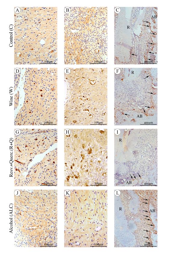
36

FIGURE 4 - Photomicrographs showing the immunostaining for OPG (A, D, G, J), IL-10 (B, E,
H, K), TRAP (C, F, I, L) in periapical periodontitis around the foraminal opening of the distal root
of the lower first molar in Control group (A, B, C), Wine group (D, E, F), Resveratrol+Quercetin
group (G, H, I), and Alcohol group (J, K, L). Counterstaining: Harris haematoxylin. Original
magnification for OPG and IL-10: 400X with scale bars: 100 μm, and 100X for TRAP with scale
bars: 400 μm. Regarding OPG and IL-10 note a large amount of immunoexpression of these
proteins in the R+Q group. For TRAP, black arrowheads points to osteoclastic cells reabsorbing
the alveolar bone (AB) located below the opening of the apical foramen. Note a reduced TRAP
immunolabeled cells in the R+Q group.
37

DISCUSSION

       Red wine or its polyphenols compounds are consumed by practically the entire world
population, regardless of the source. The present study is the first one evaluating the effect of red
wine consumption or the association between resveratrol and quercetin through oral gavage on the
development of periapical periodontitis in rats, which revealed a beneficial effect of isolated
consumption of these polyphenols on the inflammatory process magnitude and bone resorption
associated with periapical periodontitis, and the red wine consumption led to less osteoclastic
marking than the control, rejecting the null hypothesis. Although light to moderate wine intake
seems to have some beneficial effects, there is still a long way to go before definitive
recommendations on wine intake can be made (Minzer et al. 2020). Furthermore, excessive alcohol
consumption represents a public health concern (Schuckit 2009).

       The administration model via oral gavage in rats is widely used, since it allows the precise
control of the compound amount to be ingested by the animal, differently from the free availability
in the water source (Venturini et al. 2010, Correa et al. 2018). Also, in human studies, the
polyphenols investigated may come from countless types of food. Regarding wine consumption, is
difficult to determine and remain constant throughout the experimental time, since they are based
on the collaboration of the patient regarding the protocol imposed for prospective studies and the
accuracy of the answers provided in cases of retrospective studies. The 4.28 mL/kg of body weight
dosage of red wine given to the animal in the present study was used based on the general
recommendation of consumption of 300 mL/day of red wine for humans weighing seventy
kilograms (Rotondo et al. 2001, Pavlidou et al. 2018). In order to have a homogeneous comparison,
the wine used in our investigation was first subjected to the quantification of the polyphenols of
interest, through an established methodology (HPLC / DAD / ESI-MS) (Nixdorf & Hermosin-
Gutierrez 2010, Lago-Vanzela et al. 2011a, Lago-Vanzela et al. 2011b). This quantification
allowed the group receiving the two associated polyphenols to receive the exact same amount that
would be present in the wine dose administered to animals in group W.

       The infection induced model through the exposure of the rat’s pulp molars was chosen due
to the similarity of periapical response to pulp exposure as those seen in humans, once the bacterial
infection from the oral environment infects the pulp tissue leading to pulp necrosis, resulting in PP
38
(Yamasaki et al. 1994). The thirtieth-day post-lesion induction is considered an adequate time to
assess the extent of the inflammatory process and bone resorption resulting from the injury, once
the plateau stage of development has already been reached (Yamasaki et al. 1994).
The histological analysis showed an inflammatory process associated with PP of lesser
extent and intensity in the R+Q group when compared to the other groups. This finding is in
agreement with those already published in the literature proving the anti-inflammatory activity of
the two polyphenols. Resveratrol, for instance, protects from inflammation by acting at different
phases of inflammation; this anti-inflammatory activity may be performed through the inhibition
of both cyclooxygenase 1 (COX-1) and cyclooxygenase 2 (COX-2) mediated pro-inflammatory
signaling, suppression of pro-inflammatory mediator production, acting in nuclear transcription
factor-kB (NFkB) in macrophage inhibition, suppressing interleukin-6 release and interleukin-8,
which plays an important role in inflammation as it recruits leucocytes to the lesion area (Das &
Das 2007). Quercetin, in turn, downregulates nitric oxide synthase (NOS) expression, inhibit
matrix metalloproteinases, inhibits the production of inflammation-producing enzymes (COX) and
lipoxygenase (LOX), blocks TNF-α, and prevents it from directly activating extracellular signal-
related kinase (ERK) and (NF-kB), which are potent inducers of inflammatory gene expression and
protein secretion (Li et al. 2016).
The alcoholic solution in the 12.5% concentration administered alone presented the worst
results, thus excluding the hypothesis that low alcoholic levels could be one of the factors
responsible for the beneficial effects of red wine consumption. Results that agree with those already
published in the literature, which showed a deleterious effect of 15% and 20% alcohol
concentration in PP (Dal-Fabbro et al. 2019a, Dal-Fabbro et al. 2019b). Although the red wine
given in the present study has the same alcohol concentration, the inflammatory infiltrate was
significantly lower when comparing these two groups. A possible explanation is that the
nonalcoholic compounds present in the red wine, such as polyphenols, may reversed the harmful
effect evoked by the alcoholic concentration present in the red wine (de Lorimier 2000).
Even though in the present study no significant reduction in IL-1β and TNF-⍺ levels was
achieved through red wine consumption or polyphenols therapy, a propensity to a higher inhibition
of these cytokine was observed in these two groups. However, numerous studies confirmed that39
resveratrol and quercetin can suppresses the TNF-⍺ release (Takada et al. 2004, Lee & Moon 2005,
Yuan et al. 2018, Lee et al. 2019, Chen et al. 2020, Wang et al. 2020). TNF-α is a proinflammatory
cytokine released by macrophages and presents vital role in periodontitis mediated bone loss by
inducing the expression of mediators that amplify or sustain the inflammatory response such as
prostaglandins and matrix metalloproteinases (Graves & Cochran 2003). Also, IL-1 and TNF-α
acts synergistically enhancing bone resorption, and TNF play a critical role in to prepare the innate
host response to defend against bacteria, except when overstimulated, which can cause significant
collateral damage (Graves & Cochran 2003). Moreover, the pro-inflammatory cytokine IL-1β,
primarily secreted by macrophage, is a key regulator of host responses to microbial infection, being
frequently found in elevated levels in persistent periapical periodontitis (Yang et al. 2018). IL-1β
is a potent stimulator of periodontal tissue breakdown, and its properties includes promotion of
bone resorption and production of tissue-degrading proteinases (Cheng et al. 2020). A possible
explanation for the lack of significance in the reduction found in our study may be due the dosage
of resveratrol and quercetin administered, since they were lower than those applied by other studies
that showed the effectiveness in reducing this interleukin (Napimoga et al. 2013, Ribeiro et al.
2017).
Moreover, the supplementation with the associated resveratrol and quercetin caused the
elevated release of IL-10 in a significant way. Red wine, in turn, also increased the production of
IL-10, but without significance, probably due to the presence of alcohol in the drink, hindering the
beneficial effects of polyphenols (Gavala et al. 2015). IL-10 is a pleiotropic cytokine with potent
anti-inflammatory ability that suppresses both immunoproliferative and inflammatory responses,
regulates B-cell proliferation and differentiation, and downregulates vast processes such as the
release of proinflammatory cytokines and chemokines, such as IL-1, IL-6, and TNF-α, the
production of nitric oxide, and collagenase (Armstrong et al. 1996, Sun et al. 2019). Moreover, IL-
10 affects osteoclast precursors, and inhibits osteoclast activation and has been regarded as a key
regulator of bone homeostasis, in homeostatic and inflammatory conditions, once the IL-10 lack in
animals leaded to increased femur and alveolar bone loss (Cheng et al. 2020). Besides that, Il-10
is recognized as an important suppressor factor for periodontal disease and apical periodontitis
development in vivo (Sasaki et al. 2000, Sasaki et al. 2004). The molecular mechanism of bone
loss prevention evoked by the IL-10 is based on the upregulation in OPG expression and
downregulation expression of the RANKL (Cheng et al. 2020).40
The bone tissue is a dynamic complex that constantly undergoes renovation and repair (or
remodeling). The cells responsible for this process are osteoblasts, which secrete new bone, and
osteoclasts that remove the old ones. Normally, the fine balance between these two cells is in
harmony, so there is no increase or loss of bone mass. The control of bone metabolism is a key
factor in obtaining a reduction in bone resorption in inflammatory diseases (Hadjidakis &
Androulakis 2006). Therefore, the use of substances that are capable of interfering positively in
inflammatory processes that lead to bone resorption should be considered.
A number of signaling pathways maintain the activities of osteoblasts and osteoclasts; one
of the most important and frequently targeted as a new treatment strategy in bone related-disease
conditions is the RANK/RANKL/OPG system (Silva & Branco 2011). The OPG/RANKL ratio is
considered an important information to assess the cellular state of bone tissue, since the OPG is an
osteoprotective protein, acting by binding to RANKL preventing it from binding to RANK and
sequencing osteoclastic formation (Silva & Branco 2011). In the present study, as the OPG-
RANKL pathway was shifted towards OPG in group R+Q, due to a higher expression of this
protein and no differences in RANKL, less formation of bone resorptive cells occurred. This data
is confirmed by the decreased number of positive TRAP multinucleated cells (osteoclasts) per
millimeter of PP perimeter in the same group. Reduced periapical bone destruction was evidenced
by the μCT analysis in the R+Q group reinforcing the correlation between reduced TRAP-positive
cells found on immunohistochemistry, displaying attenuated bone loss due to the lower osteoclast
activities in the PP region when compared to control. Red wine consumption also led to less TRAP
multinucleated cells when compared to control, but this finding did not reflect in a smaller volume
lesion analyzed by μCT. Previous studies reinforce the action of resveratrol and quercetin on OPG
(Ribeiro et al. 2017, Ge et al. 2020). In addition to this pathway, others not evaluated in the present
study, but already published in other areas, may be related to this diminished bone resorption
presented by the phenolic group (Wattel et al. 2004, He et al. 2010). The group receiving alcohol
alone showed the highest number of TRAP-positive cells per millimeter of the PP perimeter,
confirming the deleterious effect of alcohol consumption on this marker in the PP induced in rats
(Dal-Fabbro et al. 2019a, Dal-Fabbro et al. 2019b). Interestingly, when comparing this group with
wine, the latter one had a significantly lower cell count, leading us to believe that the polyphenols
present in the drink counterbalanced the deleterious effect of alcohol.41
A prospective cohort study found that intake of wine is inversely associated with clinical
attachment loss in men (Kongstad et al. 2008). Another study in southern Brazilian adults showed
evidence of a beneficial effect of wine on periodontal status (Susin et al. 2015). Previous
investigations in ligature-induced periodontitis in animals pointed to promising paths regarding the
use of the polyphenols present in the red wine. Firstly, continuous administration of resveratrol
decreased periodontal breakdown induced experimentally in rats (Casati et al. 2013). Resveratrol
administered via oral gavage to rats caused a significant reduction in inflammation-mediated
destruction of periodontal soft tissues and bone (Correa et al. 2017). When given by subcutaneous
injection in the same lesion model, it protected rats from periodontal tissue damage by inhibiting
inflammatory responses and by stimulating antioxidant defense systems (Bhattarai et al. 2016).
The same results observed when administered freely in drinking water (Tamaki et al. 2014).
Moreover, resveratrol had a positive influence in decreasing periodontal breakdown during
smoking in rats (Ribeiro et al. 2017). Similar to resveratrol, quercetin exhibited protective effects
in bacterial-induced periodontitis, reducing the alveolar bone loss by mechanisms involving the
reduction of pro-inflammatory cytokine production and down-regulation of the osteoclastogenic
cytokine RANKL (Napimoga et al. 2013). In addition, it reduced alveolar bone loss in ligature-
induced periodontitis by increasing osteoblastic activity, decreasing osteoclastic activity,
apoptosis, and inflammation (Taskan & Gevrek 2020). These data, concomitantly with the findings
in our study, spotlight a promising approach to inhibit the development of bone loss during
periapical periodontitis development.
Considering the high prevalence of PP throughout life, combined with frequent intake of
red wine and the polyphenols through other sources, the present study offers some insights
regarding the mechanisms of how these compounds may affect the periapical periodontitis which
had not been addressed in the endodontic literature. However, due to some limitations such as the
use of an animal model, the resveratrol and quercetin dosage, the ingestion frequency, and time of
administration treatment before the periapical injury induction, the results cannot be extrapolated,
and therefore, more studies are encouraged considering these relevant parameters.42
CONCLUSION
Red wine administration led to lowers PP inflammation, TRAP marking, and periapical
bone resorption compared to ALC; resveratrol-quercetin administration reduced the PP
inflammatory processes, periapical bone resorption, and altered the OPG, IL-10, and TRAP
expression compared to C and ALC groups.
DISCLOSURE STATEMENT
The authors have stated explicitly that there are no conflicts of interest in connection with
this article.43 REFERENCES Armstrong L, Jordan N, Millar A (1996) Interleukin 10 (IL-10) regulation of tumour necrosis factor alpha (TNF-alpha) from human alveolar macrophages and peripheral blood monocytes Thorax 51, 143-9. Artero A, Artero A, Tarín JJ, Cano A (2015) The impact of moderate wine consumption on health Maturitas 80, 3-13. Astolphi RD, Curbete MM, Colombo NH et al. (2013) Periapical lesions decrease insulin signal and cause insulin resistance Journal of Endodontics 39, 648-52. Azuma MM, Gomes-Filho JE, Ervolino E et al. (2017) Omega 3 Fatty Acids Reduce Bone Resorption While Promoting Bone Generation in Rat Apical Periodontitis Journal of Endodontics 43, 970-6. Bhattarai G, Poudel SB, Kook SH, Lee JC (2016) Resveratrol prevents alveolar bone loss in an experimental rat model of periodontitis Acta Biomater 29, 398-408. Boots AW, Haenen GR, Bast A (2008) Health effects of quercetin: from antioxidant to nutraceutical European Journal of Pharmacology 585, 325-37. Casado-Díaz A, Anter J, Dorado G, Quesada-Gómez JM (2016) Effects of quercetin, a natural phenolic compound, in the differentiation of human mesenchymal stem cells (MSC) into adipocytes and osteoblasts The Journal of Nutritional Biochemistry 32, 151-62. Casati MZ, Algayer C, Cardoso da Cruz G et al. (2013) Resveratrol decreases periodontal breakdown and modulates local levels of cytokines during periodontitis in rats Journal of Periodontology 84, e58-64. Chen T, Zhang X, Zhu G et al. (2020) Quercetin inhibits TNF-α induced HUVECs apoptosis and inflammation via downregulating NF-kB and AP-1 signaling pathway in vitro Medicine (Baltimore) 99, e22241. Cheng R, Wu Z, Li M, Shao M, Hu T (2020) Interleukin-1β is a potential therapeutic target for periodontitis: a narrative review International Journal of Oral Science 12, 2. Cintra LT, da Silva Facundo AC, Prieto AK et al. (2014) Blood profile and histology in oral infections associated with diabetes Journal of Endodontics 40, 1139-44. Cintra LT, Samuel RO, Azuma MM et al. (2016) Multiple Apical Periodontitis Influences Serum Levels of Cytokines and Nitric Oxide Journal of Endodontics 42, 747-51. Correa MG, Pires PR, Ribeiro FV et al. (2018) Systemic treatment with resveratrol reduces the progression of experimental periodontitis and arthritis in rats PloS One 13, e0204414. Correa MG, Pires PR, Ribeiro FV et al. (2017) Systemic treatment with resveratrol and/or curcumin reduces the progression of experimental periodontitis in rats Journal of Periodontal Research 52, 201-9. Cosme-Silva L, Dal-Fabbro R, Cintra LTA et al. (2019) Systemic administration of probiotics reduces the severity of apical periodontitis International Endodontic Journal 52, 1738-49.
También puede leer