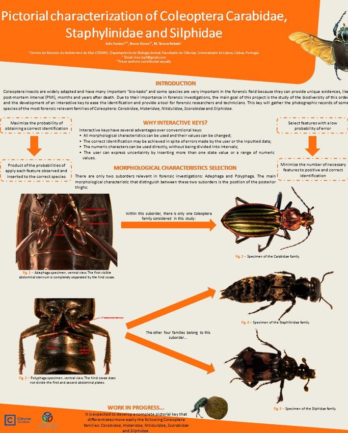
Pictorial Characterization of Eight Coleoptera Families with Forensic Interest
←
→
Transcripción del contenido de la página
Si su navegador no muestra la página correctamente, lea el contenido de la página a continuación
UNIVERSIDADE DE LISBOA
                     FACULDADE DE CIÊNCIAS
               DEPARTAMENTO DE BIOLOGIA ANIMAL

Pictorial Characterization of Eight Coleoptera Families with
                      Forensic Interest

             Inês de Lima e Santos Pimentel Fontes

             Mestrado em Biologia Humana e Ambiente

                      Dissertação orientada por:
               Professora Doutora Maria Teresa Rebelo

                               2016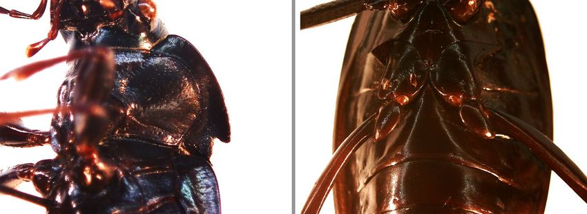
UNIVERSIDADE DE LISBOA
                   FACULDADE DE CIÊNCIAS
          DEPARTAMENTO DE BIOLOGIA ANIMAL

   Pictorial Characterization of Eight Coleoptera
           Families with Forensic Interest

                Inês de Lima e Santos Pimentel Fontes

                Mestrado em Biologia Humana e Ambiente

                               Dissertação

                   Dissertação orientada por:
Prof. Doutora Maria Teresa Rebelo, Departamento de Biologia Animal,
Faculdade de Ciências da Universidade de Lisboa, 1749-016 Lisboa, Portugal

                                  2016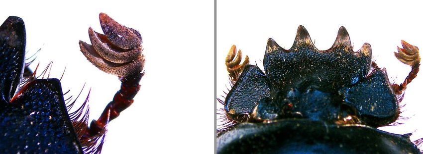
Dissertação para a obtenção do
grau de mestre em Biologia
Humana e Ambiente.

Trabalho realizado no Laboratório
de Entomologia (Departamento de
Biologia Animal) da Faculdade de
Ciências da Universidade de
Lisboa.
Previous Note
The pictorial characterizations presented in this work are still under construction. Species
collected in future experiments can be added to this key in order to help a better
characterization of Portuguese Coleoptera fauna.

The results of the present work lead to the creation of pictorial keys and characterizations
available online (www.csicoleoptera.weebly.com).

It was submitted to an online scientific journal (Ecologi@) an article with the preliminary
results of this project and these same results were presented at 2015 Encontro sobre
Biodiversidade e Conservação de Invertebrados in a scientific poster.

The references used in this dissertation are in accordance with the norms of the journal
Forensic Science International.
One day Alice came to a fork in the road and saw a Cheshire cat in a tree.
                                                                     ‘Which road do I take?’ she asked.
                                                         ‘Where do you want to go?’ was his response.
                                                                        ‘I don’t know,’ Alice answered.
                                                                 ‘Then,’ said the cat, ‘it doesn’t matter.’

                                                 Lewis Carroll, Alice's Adventures in Wonderland (1865)

                                 The very basic core of a man's living spirit is his passion for adventure.
     The joy of life comes from our encounters with new experiences, and hence there is no greater joy
              than to have an endlessly changing horizon, for each day to have a new and different sun.
If you want to get more out of life, you must lose your inclination for monotonous security and adopt a
                                     helter-skelter style of life that will at first appear to you to be crazy.
 But once you become accustomed to such a life you will see its full meaning and its incredible beauty.

                                                                         Jon Krakauer, Into the Wild (1996)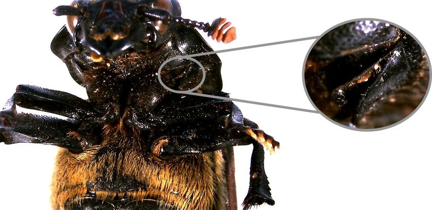
AGRADECIMENTOS
   Para grande felicidade minha, percebi que o esforço necessário à elaboração desta tese de mestrado
não foi apenas individual mas também proveio do apoio e incentivo de um conjunto de pessoas às
quais devo o meu mais sincero agradecimento.

   Quero agradecer à Professora Doutora Maria Teresa Rebelo, minha oridentadora, pelo apoio,
palavras de incentivo e boa disposição sempre presente.

   A todo o pessoal da Sociedade Portuguesa de Entomologia e, em especial, à Carla e ao Mário pela
boa disposição em todas as atividades que participámos, desde os mercados no museu à organização
do congresso ibérico de entomologia. Ao Telmo Nunes, pela simpatia e disponibilidade para me
ensinar a trabalhar com o estereomicroscópio e Photoshop. À Sílvia Diniz e à Carla Loução da
Faculdade de Medicina Veterinária pela cedência de alguns exemplares e ao Professor Artur Serrano
pelo empréstimo do espécime de Scarabaeidae com as antenas lameladas mais perfeitas.

   Aos meus pais, que sempre me transmitiram os valores do amor e respeito e forneceram os
fundamentos morais e éticos que guiam a minha vida e me permitem valorizar o conhecimento. Mãe,
podes continuar a contar as tuas histórias cada vez mais ramificadas à medida que a narrativa avança
que eu vou estar sempre aqui para ouvi-las até ao fim mas não sem antes soltar uns quantos risos e
suspiros de impaciência; continua a arranjar músicas cantadas inesperadamente no fim de cada frase.
Pai, meu companheiro incansável de caminhadas e aventuras com o complicómetro sempre ligado.
Temos que começar a planear a nossa próxima grande caminhada!

   Aos meus dois maninhos mais novos, um alentejano mecatrónico com sotaque carregado e outro
massagista (quase) profissional que me aliviou os stresses, muitos deles nas semanas que precederam a
entrega da tese. Sem vocês não seria a mana mais velha e responsável (coff coff…) que sou hoje!

    Ao avô Fontes que sempre demonstrou uma paciência inabalável para todos os seus netos, sentido
criativo fora do vulgar e uma veia poética única. Já está na altura de escrever o segundo volume das
Pilhérias dos Netos!

   À avó Lena, minha professora de inglês e que é um tesouro de pessoa, cheia de força e sempre
dedicada à família. Recordarei para sempre as férias passadas na casa dos três porquinhos e as
histórias sobre a aventura noturna de recuperação do livro de orações da rainha no palácio de Vila
Viçosa.

   Em memória do avô Manel que me ajudou a gostar um bocadinho mais de química e de quem eu
sempre admirei a coragem para experimentar gastronomia diferente. De certeza que aprovaria a
entomofagia!

   Em memória da avó Ceição que foi a responsável por aguçar a minha veia forense com as histórias
do Poirot e da Miss Marple. As tardes passadas no quintal da vivenda a brincar, a comer pão com
manteiga sem côdea e a partir pinhões é uma das minhas memórias de infância mais querida.

    A todos os meus tios, tias, primos e primas, de ambos os lados, que não vou discriminar porque
felizmente somos uma família muito grande e unida. Vocês são uma fonte inesgotável de apoio e boa
disposição. Não posso deixar de agradecer às minhas tias-avós do coração, a tia Inês e a tia Naninha,
que sempre acreditaram no meu sucesso e me animaram nos almoços em Benfica.

                                                                                                  vii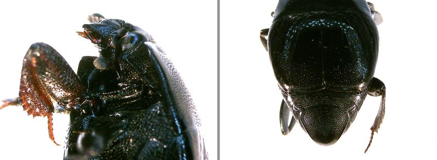
À minha mana do coração, Ana Magali, minha companhia de sereia do fontanário, que está e
sempre esteve presente. As nossas palhaçadas, roteiros gastronómicos bastante diversificados,
aventuras turísticas e cinematográficas (por piores que sejam os filmes) conseguem sempre animar os
meus dias. Obrigada por todos os risos, desabafos e todo o carinho, amizade e apoio que me dás. Vais
ver que um dia damos por nós numa praia na Nova Zelândia, ao pé de uma colmeia de formigas
afogadas, a comer uma bela melancia e a beber duas cervejas de lata, se elas estiverem capazes.

   À minha irmã Esmy. Dizem que os verdadeiros amigos se mantêm presentes mesmo estando longe
e posso dizer que és uma delas. És uma das pessoas mais fortes e determinadas que conheço. Aos
meus amigos Miguel e Paulo, os meus guarda-costas e que estão sempre presentes, mesmo nos
momentos mais difíceis.

   À Maria João, minha querida amiga que me veio lembrar que o mundo é do tamanho de um grão de
areia. A tua honestidade foi uma das primeiras coisas que conheci em ti e, a partir daí, só vieram
coisas boas. Obrigada por todo o apoio que me deste ao longo desta árdua etapa e que foi
acompanhada de uma brutalíssima banda sonora pop, assim como só nós sabemos apreciar.

   Ao meu irmão espanhol, Alberto Fuentes (És Fuentes? Eu sou Fontes! Fuentes! Fontes!), uma
pessoa espetacular e que me trouxe imensas coisas boas neste último ano. As nossas viagens de
amostragem foram, sem dúvida, o ponto alto, mesmo com as poucas horas de sono e o infindável (e
mal cheiroso) trabalho de armadilhagem. Ensinaste-me a ser turista na minha própria cidade e agora
quero que me ensines a ser turista em Caravaca e Múrcia. Obrigada por teres comido o resto da minha
francesinha, eu já não conseguia comer mais!

   À Nocas, minha irmã CSI e recente mestre. A nossa amizade já passou por muito e saiu sempre
mais forte! À melhor professora do mundo, Ana Paula Ferreira, que mostrou um empenho e dedicação
sem comparação nas melhores aulas que um aluno de ciências forenses e criminais pode ter: análise do
local do crime. Felizmente passaste de professora para amiga e sempre estiveste ao meu lado e
acreditaste em mim. Aqui a caramela fininha está à espera do próximo jantar!

   A todo o gang da Casa do Alentejo que, apesar de haver algumas novidades de vez em quando, há
uns quantos que estão sempre presentes. Ritinha, obrigada pela boa disposição original que tens
sempre para a mai nova. Sandrinha, ainda estou à procura dos pulgões e à espera da próxima festa da
marquise nas Américas!

    A todo o pessoal que conheci ao longo do mestrado e que foram ficando e, por isso, se tornaram
amigos. Em especial à Joana Ivone, the mother of spoilers e telefonista da segurança social, por todas
as parvoíces, maluquices e peixeiradas que fazemos sempre que estamos juntas; à Madalena, à
Daniela, à Naila e ao Bruno pela vossa companhia mais ou menos regular nas horas de almoço, cafés e
momentos de pura procrastinação dos quais vocês são os únicos culpados. Ao Flávio stufu, meu
companheiro de gabinete, pela paciência que tens em ouvir as minhas cantorias (des)afinadas. À
Carina estorninha que, apesar de já te conhecer há mais tempo, este inesperado reencontro neste
último ano trouxe-me bastante alegria.

   Por fim, quero agradecer à minha família da Casa da Sorte. Patty, da difícil tarefa que tínhamos em
mãos todos os dias, felizmente conseguimos formar uma grande amizade. Sr. Vítor, parece que é desta
que entrego a tese mas não se preocupe que assim que o fizer deixo o meu currículo aí outra vez.
Mário padrinho, eu sei que nunca lhe vendi a sorte grande mas esta amizade é muito mais valiosa!

   Sem a vossa força e motivação não teria conseguido!

                                                                                                  viii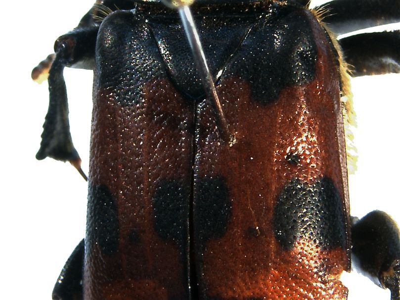
SUMÁRIO
   Desde tempos remotos, os insetos têm suscitado um interesse no ser humano, o que se pode
confirmar através da observação de artefactos de civilizações antigas como os Egípcios, Chineses,
Maias e Aztecas. Estas obras de arte, como pinturas e esculturas, frequentemente associavam
determinados insetos não só à morte mas também à reincarnação e vida pós-morte.

   A aplicação de conhecimentos entomológicos em investigações criminais tem uma história bastante
extensa e bem documentada. Começando no primeiro caso registado da aplicação da entomologia na
resolução de um homicídio na China Medieval, a utilização desta ciência como uma ferramenta
forense tem vindo a percorrer um longo caminho repleto de contribuições científicas provenientes dos
quatro cantos do mundo. No entanto, foi a investigação do homicídio de um recém-nascido em França
no final do século XVII que marcou o início da entomologia forense moderna e que contribuiu para a
publicação da notável obra de Mégnin “La Faune des Cadavres: Application de l’Entomologie à la
Médicine Légale”. A partir de então, esta ferramenta forense foi desenvolvida principalmente através
de contribuições de investigações científicas durante os anos das grandes guerras e a sua completa
aceitação só foi possível devido ao trabalho conjunto de académicos e forças de segurança.

   Os insetos podem fornecer importantes informações em investigações forenses não só em casos de
homicídios como também em casos de negligência tanto de seres humanos como de animais
domésticos ou selvagens, por exemplo, em jardins zoológicos. Adicionalmente, numa área
complementar designada entomotoxicologia, conjugam-se os conhecimentos da entomologia e da
toxicologia para responder a diversas questões, como a possibilidade da presença de substâncias
tóxicas no cadáver, em casos de homicídio, suicídio ou mortes acidentais. A entomologia forense pode
igualmente ser utilizada em casos de investigação de pragas tanto num âmbito imobiliário como
alimentar.

   Contudo, a utilização dos conhecimentos entomológicos em investigações de homicídios é,
possivelmente, a mais mediatizada, principalmente pelos meios de comunicação social e
cinematográficos. Uma vez que contribuem para a decomposição do cadáver, os insetos constituem
evidências cruciais. Assim sendo, quando é descoberto um cadáver, os insetos possibilitam o
conhecimento de diversos parâmetros que, em certos casos, seriam impossíveis de descobrir utilizando
outros campos do saber, nomeadamente as técnicas médico-legais, como é o caso da temperatura
corporal. Os insetos que colonizam o cadáver permitem não só estimar o intervalo pós-morte (IPM)
através dos seus padrões de desenvolvimento e sucessão, como também poderão auxiliar na descoberta
do local do crime primário nos casos em que há a suspeita de que o cadáver foi movido postmortem.
Contudo, a primeira grande etapa é a recolha, no local do crime, de todos os vestígios entomológicos e
sua correta identificação. No entanto, este potencial forense dos insetos só pode ser totalmente
reconhecido se se realizarem extensos estudos a fim de compreender melhor não só a sua biologia e
funcionamento enquanto seres vivos, como também a sua bioecologia, ou seja, as interações que estes
realizam com o meio à sua volta. Contudo, a aplicação de tais estudos a investigações é um processo
que necessita de ser realizado com caução uma vez que, estando distribuídos globalmente, os insetos
apresentam diferenças de comportamento. Adicionalmente, existem fatores que são relevantes e que
necessitam de uma cuidada análise, como é o caso das condições meteorológicas. Sabe-se que a
temperatura e humidade são importantes fatores que determinam a velocidade de desenvolvimento dos
insetos.

  O processo de decomposição, apesar de ser uma ocorrência contínua no tempo, pode ser dividido
em fases que são marcadas por diferenças significativas que ocorrem no cadáver. Esta divisão, que

                                                                                                    ix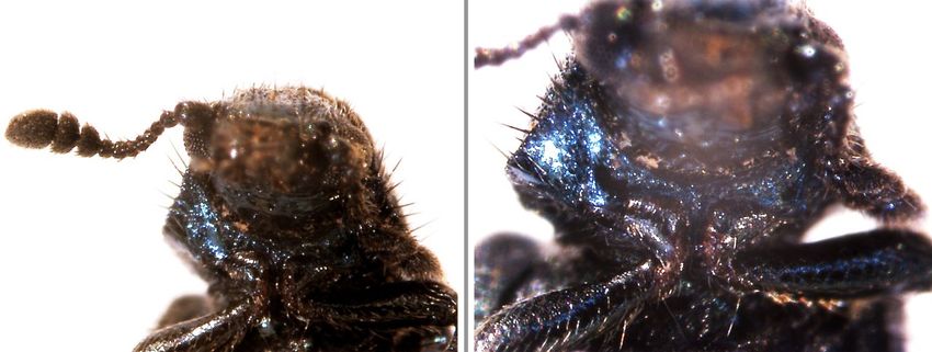
facilita a sua análise, é, por vezes, realizada diferentemente consoante o investigador mas,
frequentemente, considera-se que todo o processo de decomposição é composto por cinco fases
diferentes: fase fresca, fase de inchaço, fase de decomposição ativa, fase de decomposição avançada e,
por fim, a fase seca. Estas etapas são frequentemente protagonizadas por um conjunto de fauna
entomológica que auxilia a investigação e que, nos casos em que a morte ocorreu há mais de 72 horas,
tornam-se evidências cruciais.

    Apesar de muitos insetos visitarem cadáveres em decomposição, as ordens que mais relevância têm
no âmbito da entomologia forense são a Diptera (moscas) e a Coleoptera (escaravelhos). As moscas
são conhecidas por serem os primeiros colonizadores de cadáveres e são, sem dúvida, as mais
utilizadas em casos em que conhecimentos de entomologia forense são requeridos.

    Ao chegarem ao cadáver, depositam os seus ovos que, após eclodirem, originam larvas que se vão
alimentar dos tecidos em decomposição. Estas passam por várias fases larvares, demarcadas pelas
diferenças em tamanho, e, quando prontas, pupam por um período variável de tempo. Quando
completo o processo de metamorfose dentro da câmara de pupação, adultos emergem, prontos para
iniciar um novo ciclo. Uma das técnicas mais utilizadas em entomologia forense é a utilização das
fases de desenvolvimento larvar das moscas para a estimativa do IPM. Este método tem, no entanto,
uma série de condicionantes que necessitam de cuidada atenção, como é o caso da temperatura e
humidade a que as larvas estão expostas. Estas variáveis podem alterar significativamente o tempo de
desenvolvimento uma vez que estes insetos são poiquilotérmicos, condicionando assim a estimativa do
IPM.

    Outro método utilizado para estimar o IPM é a sucessão entomológica de insetos no cadáver.
Sabendo que diferentes fases de decomposição atraem diferentes espécies de insetos, a utilização deste
padrão único de sucessão pode ser bastante útil, especialmente em casos em que o IPM é mais longo.
O trabalho pioneiro de Mégnin, que descreveu a existência de oito ondas de colonização de cadáveres
ao longo das cinco fases de decomposição, é ainda nos dias de hoje, amplamente referido quando se
utiliza esta técnica. Em casos em que é necessário utilizar estes conhecimentos o cadáver já se
encontra, frequentemente, em estados de decomposição mais avançados e as moscas deixaram de ser
os seus principais colonizadores, passando a ser os escaravelhos os artrópodes mais relevantes. Apesar
de poderem estar presentes nas fases iniciais de decomposição (frequentemente como predadores de
fases larvares de outros insetos), os escaravelhos encontram-se mais associados a fases tardias deste
processo e, muitas vezes, as únicas evidências entomológicas em casos nas quais a morte ocorreu há
meses ou até anos.

   Apesar de ser uma ferramenta válida e estabelecida no âmbito das ciências forenses, a entomologia
forense é ainda pouco utilizada em Portugal. No entanto, a realização de estudos científicos cujo
principal objetivo é a compreensão das interações entre insetos e cadáveres tem sido uma importante
contribuição para o desenvolvimento desta ciência em Portugal.

    Este estudo centra-se em oito famílias de coleópteros presentes em Portugal, fortemente
relacionadas com casos de entomologia forense: Carabidae, Cleridae, Dermestidae, Histeridae,
Nitidulidae, Scarabaeidae, Silphidae e Staphylinidae. O seu principal objetivo é utilizar imagens para
caracterizar morfologicamente as espécies mais representativas dessas famílias. Para tal, foram
utilizados espécimes previamente capturados em armadilhas com isco ou em carcaças de animais
provenientes de diversas zonas de Portugal: Serra da Estrela, Sertã, Campo Grande e Aroeira. Estes
espécimes foram recolhidos utilizando armadilhas do tipo “pitfall” e “malaise” e, após a sua captura.,
preservados em álcool 70%. Seguidamente, montaram-se os exemplares em alfinetes entomológicos e

                                                                                                    x
procedeu-se à sua observação utilizando um estereomicroscópio. Com o auxílio de chaves dicotómicas
e galerias fotográficas, procedeu-se à sua identificação e seleção de características diagnosticantes que
foram posteriormente fotografados num segundo estereomicroscópio com melhor definição. O registo
fotográfico obtido foi então utilizado para a construção de chaves pictóricas interativas e
caracterizações fotográficas, disponíveis online (www.csicoleoptera.weebly.com). Esta é uma
importante ferramenta de auxílio em investigações com os mais variados quesitos uma vez que
permitirá uma fácil e rápida identificação de grupos relevantes por técnicos forenses sem experiência
em entomologia.
Palavras-chave: Entomologia forense, Coleoptera, características diagnosticantes, chaves de
identificação, microscopia ótica.
xiABSTRACT
Since ancient times, humans have shown a unique interest in insects, which can be seen in artworks
of ancient civilizations like the Egyptians, Chinese, Mayans and Aztecs. These works of art, like
paintings and sculptures, often associate certain insects not only with death but also with the
reincarnation and afterlife.
Despite of the fact that criminal investigations arouse the most curiosity within the scope of
forensic entomology, this science can be used to deal with a wide range of other investigations.
Neglect cases in civil investigations are also known to use this tool to answer several questions.
Another known and well developed area is the entomology of stored products. In the last years, the
union of toxicology and entomology created another field of work within forensic entomology:
entomotoxicology. This recent tool is very useful to know if, at the time of death, toxic substances
were present in the body.
Insects can provide significant information in death investigations because of their contribution to
corpse decomposition. As so, if a dead body is discovered, insects can help in the estimation of the
postmortem interval (PMI). However, to apply entomological knowledge to criminal investigations is
essential not only to have information regarding insects’ bioecology that is valid for the geographic
place where the body was found but also to accomplish an accurate identification of the species found
on the corpse.
The focus of this study lies in eight Coleoptera families present in Portugal, strongly related to
forensic entomology cases: Carabidae, Cleridae, Dermestidae, Histeridae, Nitidulidae, Scarabaeidae,
Silphidae and Staphylinidae. Its main goal is to use pictures to characterize morphologically the most
representative genus and species belonging to these families using specimens previously caught in
baited traps or on animal carcasses. To proceed with this analysis, the diagnostic structures of the
specimens are described and photographed using optical microscopy and the data obtained was used to
build pictorial interactive keys and characterizations, available online (www.csicoleoptera.
weebly.com). This will allow an easy identification of relevant groups for forensic technicians without
expertise in entomology.
Keywords: Forensic entomology, Coleoptera, diagnostic structures, identification keys,
microscopy.
xiiTABLE OF CONTENTS
Agradecimentos ...................................................................................................................... vii
Sumário ..................................................................................................................................... ix
Abstract .................................................................................................................................... xii
Table of Contents ................................................................................................................... xiii
List of Figures ......................................................................................................................... xv
List of Tables ........................................................................................................................... xviii
List of Abbreviations ............................................................................................................. xix
I. State of the Art ................................................................................................................... 1
1. Forensic Entomology ..................................................................................................... 2
1.1. Definition and main areas ....................................................................................... 2
1.2. History ......................................................................................................................... 2
1.2.1. Historical records of early human civilizations ........................................................
2
1.2.2. Medieval China, artworks and early influences ........................................................ 3
1.2.3. Casework, research, war and public policy that made the groundwork of forensic
entomology ............................................................................................................... 3
1.2.4. The twentieth century: baby deaths investigations contributed to new advances in
understanding necrophagous insects ......................................................................... 5
1.2.5. Forensic entomology during the World Wars ........................................................... 6
1.2.6. Developments after the “great” Wars ....................................................................... 7
1.2.7. Recent history and the growth of forensic entomology ............................................ 7
2. Medico-legal Entomology ............................................................................................. 8
2.1. Postmortem changes .................................................................................................. 8
2.1.1. First postmortem changes ………………………………………………................. 8
2.1.2. Stages of decomposition ………………………………………………................... 9
2.2. Insects with forensic importance ………………………………………………... 11
2.2.1. Classification ………………………………………………………………………. 11
2.2.2. Collection of entomological evidence ……………………………………………..
13
2.3. Estimating the postmortem interval ……………………………………………... 14
2.4. Medico-legal entomology in Portugal …………………………………………... 17
3. The Coleoptera Order ………………………………………………………………... 18
3.1. General morphological characteristics…………………………………………. 18
3.2. Family characteristics …………………………………………………………….. 20
3.2.1. Carabidae .…………………………………………………………………………. 20
xiii3.2.2. Cleridae ………………………………………………………………………….… 21
3.2.3. Dermestidae ……………………………………………………………………….. 21
3.2.4. Histeridae ………………………………………………………………………….. 22
3.2.5. Nitidulidae …………………………………………………………………………. 22
3.2.6. Scarabaeidae ………………………………………………………………………. 23
3.2.7. Silphidae …………………………………………………………………………... 23
3.2.8. Staphylinidae ………………………………………………………………………. 23
4. Interactive Keys …………………………………………………………………. 24
II. Thesis Context and Objectives ...……………………………………………………… 25
III. Materials and Methods ………………………………………………………………. 27
1. Sampling ………………………………………………………………………………... 28
2. Assembly and identification …………………………………………………………. 28
3. Photographic record and key elaboration ………………………………………… 29
IV. Results and Discussion ...……………………………………………………………... 30
1. Key to Eight Coleoptera Families with Forensic Interest …...………………….. 31
2. Silphidae Key to Three Species of Nicrophorus Genus ……………………......... 39
3. Pictorial Characterization of Carabus Genus …………………….......................... 44
4. Dermestes frischii Pictorial Characterization …………………….......................... 45
5. Saprinus subnitescens Pictorial Characterization ………………………............... 47
6. Onthophagus joannae Pictorial Characterization ………………………............... 49
7. Creophilus maxillosus Pictorial Characterization …………………….................. 51
V. Conclusion and Perspectives ……………………..................………………………… 55
VI. References ……………………..................………………………................................. 57
VII. Appendices ……………………..................………………………............................... 68
1. Article submitted to online scientific journal Ecologi@ ........................................ 69
2. Poster presented at EBCI 2015 ................................................................................... 70
3. Website CSI Coleoptera ………………………………………………………… 71
xivLIST OF FIGURES
Figure 1. Decomposition stages and its relation with insect activity……………………………... 10
Figure 2. Coleoptera general morphological characteristics……………………………………… 20
Figure 3. Morphological characteristics that distinguish between Adephaga and Polyphaga:
A – presence of notopleural suture; B – posterior border of the metacoxae passes the first
abdominal sternite; C – absence of notopleural suture; D – posterior border of the metacoxae
does not pass the first abdominal sternite ………………………………………………………… 32
Figure 4. Morphological characteristics of Staphylinidae: A – lamellate antennae; B – large
clypeus covering labrum in dorsal view…………………………………………………………… 32
Figure 5. Different types of antennae: A – Carabidae filiform antennae; B – Silphidae slightly
club antennae; C – Histeridae elbowed antennae; D – Nitidulidae distinctly capitate antennae…. 33
Figure 6. Morphological characteristics of Staphylinidae: A – elytra very short and truncate
exposing more than three abdominal tergites; B – antennae filiform or moniliform……………... 34
Figure 7. Morphological characteristics of Nitidulidae and Histeridae: A – antennae with a
compact club; B – short and truncate elytra exposing abdominal tergites………………………... 34
Figure 8. Procoxae transverse with exposed trochantin on Nitidulidae………………………….. 35
Figure 9. Morphological characteristics of Histeridae: A – procoxae transverse without exposed
trochantin; B – short and truncate elytra exposing pygidium and propygidium…………………... 35
Figure 10. Tibiae flattened with spines on Histeridae…………………………………………….. 36
Figure 11. Morphological characteristics of Silphidae: A – capitate antennae with 10 or 11
segments; B – elytra with orange markings and longitudinal striae; C – head prognathous and
pronotum tomentose……………………………………………………………………………….. 36
Figure 12. Exposed trochantin on Silphidae……………………………………………………… 37
Figure 13. Head hypognathous……………………………………………………………………. 37
Figure 14. Dermestidae morphological characteristics: A – clubbed and short antennae received
into grooves on underside of prothorax; B – five visible sternites………………………………... 38
Figure 15. Body elongated with bristly hairs and pronotum narrower than elytra……………...... 38
Figure 16. Cleridae morphological characteristics: A – antennae not received into grooves on
underside of prothorax; B – conical procoxae…………………………………………………….. 38
Figure 17. Elytra appearing cut off at the apex and four abdominal segments exposed…………. 39
Figure 18. Morphological characteristics of Nicrophorus genus: A –clubbed antenna;
B – protibia with a strong tooth towards the apex………………………………………………… 39
Figure 19. Two transverse reddish to orange-brown marks present on elytra……………………. 40
Figure 20. Antennae with the last three segments of club reddish-yellow………………………... 40
Figure 21. Morphological characteristic distinguishing Nicrophorus vestigator from two other
species: A – front and rear margin of pronotum with long golden hair; B – hairless pronotum….. 41
Figure 22. Nicrophorus vestigator whole body photograph……………………………………… 41
xvFigure 23. Coloured mark from the front continues without interruption across elytra………….. 42
Figure 24. Nicrophorus investigator whole body photograph……………………………………. 42
Figure 25. Both coloured marks separated from one another at the suture……………………….. 42
Figure 26. Nicrophorus interruptus whole body photograph…………………………………….. 43
Figure 27. Morphological characteristic of Carabus spp.: A – distance between the antennae
broader than clypeus length; B – frons without ridges and with two punctures, each one carrying
a bristle; C – broad head and pronotum not oval in shape………………………………………… 44
Figure 28. Hairless elytra with (A) regular striae (less than 11) or (B) irregular sculpture with
rows of granules…………………………………………………………………………………… 44
Figure 29. Absence of waist between the pronotum and elytra…………………………………... 44
Figure 30. Carabus sp. whole body photograph………………………………………………….. 44
Figure 31. Frons without an ocellus………………………………………………………………. 45
Figure 32. Morphological characteristics of Dermestes genus: A – pronotum broadest at the
base, tapering towards the front; B – clubbed antennae; C – antenna club oval…………………. 45
Figure 33. Morphological characteristic of Dermestinus subgenus: A – semi-circular shaped
front border with right-angles; B – domed pronotum; C – underside with numerous white
hairs……………………………………………………………………………………………....... 45
Figure 34. Sides of the pronotum with white hairs pointing towards the middle………................ 46
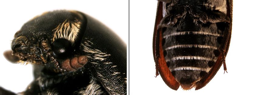
Figure 35. Dermestes frischii morphological characteristics: A – head with patches of golden
hairs; B – apex without a sharp point and with a smooth margin; C – black hair at the tip of the
abdomen’s last segment………………………………………………………................................ 46
Figure 36. Scutellum covered with yellowish hairs……………………………………………….. 46
Figure 37. Dermestes frischii whole body photograph…………………………………………… 46
Figure 38. Unruffled pronotum, broadest at the base……………………………………………... 47
Figure 39. Pronotum and elytra without uniform ridges and hairless……………………………. 47
Figure 40. Club of antenna oval and usually appearing segmented……………………………… 47
Figure 41. Gular lobe absent……………………………………………………………………… 47
Figure 42. Morphological characteristics of Saprinus genus: A – distinct border separating eyes
from frons; B – rim separating eyes from frons is discontinued…………………………………... 47
Figure 43. Morphological characteristics of Saprinus genus: A – prominent mandibles;
B – punctured elytra and with striae; C – front tibiae with teeth…………………………………. 48
Figure 44. Front half of the elytra smooth yet crossed by more than four striae, with the
innermost curving towards the elytra suture………………………………………………………. 48
Figure 45. Saprinus subnitescens morphological characteristics: A – mesosternum with the
central part without punctures; B – top of pronotum without punctures and its margin hairless;
C – stria running parallel to the rear edge connects to the sutural stria…………………………... 48
Figure 46. Saprinus subnitescens whole body photograph……………………………………….. 48
Figure 47. Elytra covering last segment of abdomen, only visible if viewed from behind;
A – dorsal view; B – view from behind; C – antennae with short club covered by minute hairs… 49
xviFigure 48. Morphological characteristics of Scarabaeinae subfamily: A – elytra as broad as
long; B – each pro tibia with a spur; C – absent scutellum………………………………………... 49
Figure 49. Morphological characteristics of Onthophagus genus: A – pronotum front margin
strongly; B – eight striae on each elytron …………………………………………………………. 49
Figure 50. Front of head sinuate in the middle and males with a transverse margin located in
front of the eyes……………………………………………………………………………………. 50
Figure 51. Onthophagus joannae whole body photograph……………………………………….. 50
Figure 52. Morphological characteristics of Staphylininae subfamily: A – elytra shortened with
more than three segments of the abdomen exposed and the last one with long styles; B – five
segmented tarsi; C – antennae not clubbed and eleven segmented, inserted under a ridge on the
front………………………………………………………………………………………………... 51
Figure 53. Morphological characteristics of Staphylininae subfamily: A – prosternum does not
project forwards under the neck; B – pronotum and elytra without ridges or longitudinal
depressions and elytra not overlapping with one another…………………………………………. 51
Figure 54. Smaller mandibles and without curved extensions and antennae inserted at the front
of the head (inside the base of the mandibles) a long way apart, further from each other than
from the eyes………………………………………………………………………………………. 51
Figure 55. Hind tarsi usually with an elongated first segment…………………………………… 51
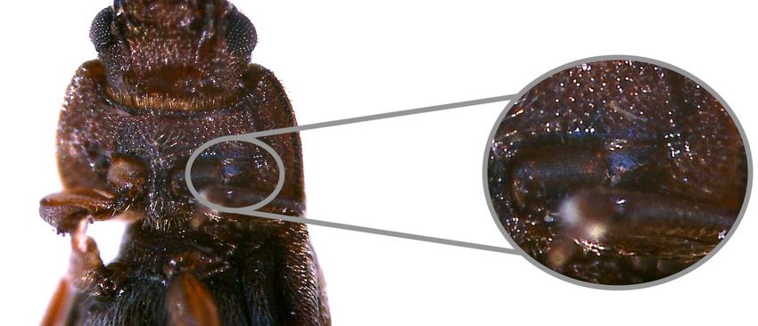
Figure 56. Morphological characteristics of Staphylinina subtribe: A – epipleura visible;
B – punctured pronotum………………………………………………………………………….... 52
Figure 57. Short antennae, longer than the head and with a club of 5 to 6 segments, broader than
long……………………………………………………………………………………………….... 52
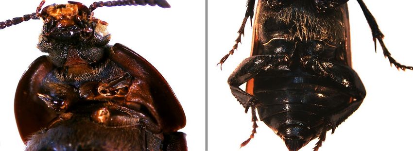
Figure 58. Morphological characteristics of Creophilus genus: A – bare head except behind the
eyes; B – bare pronotum except on the side margins and without punctures on top……………… 52
Figure 59. Distinctive yellow pubescence across middle of elytra and on abdominal tergites…... 53
Figure 60. Creophilus maxillosus whole body photograph……………………………………….. 53
xviiLIST OF TABLES
Table 1. Biotic components of carrion organisms………………………………………………… 12
Table 2. Principal members of the faunal succession on human cadavers……………………....... 15
xviiiLIST OF ABBREVIATIONS
PMI – postmortem interval
EBCI – Encontro sobre Biodiversidade e Conservação de Invertebrados
EFD – Experimental Field Devices
mag – magnification
ns – notopleural suture
mtc – metacoxae
S1 – first abdominal sternite
S2 – second abdominal sternite
nsa – notopleural suture absent
cl – clypeus
el – elytron
S1-S5 – first through fifth visible abdominal tergites
an – antenna
s – abdominal tergites
pc – procoxa
tca – trochantin absent
prp – propygidium
pyg – pygidium
eli – elytron striae
pr – pronotum
mxp – terminal palpomere of the maxillary palps
sos – supraorbital setae
fr – frons
wh – white hairs
sc – scutellum
gla – gular lobe absent
mn – mandibles
xixey – eye
bd – border
sp – spur
epi – epipleura
xxI. State of the Art
1I. State of the Art
1. Forensic Entomology
1.1. Definition and main areas
Forensic entomology is the result of the synergy between two important fields of knowledge,
forensic sciences and entomology, and therefore is where arthropod science and the legal system
cooperate [1,2]. Although most of the events where forensic entomology assists are related to cases of
homicide detected within a short period of time [1], the insects used as evidences reach far beyond just
those that exhibit necrophilic feeding habits on human cadavers [3].
In order to assist legal inquiries, it is very important to know the distribution, bioecology and
behaviour of insects found in a cadaver. This knowledge can provide relevant information to answer
the classic forensic questions about a crime: when?, where? and how? [4,5].
Although publicity surrounding forensic entomology comes from criminal cases, this branch of
forensic sciences can also assist civil inquiries [6]. In 1986, Lord and Stevenson were responsible for
the separation of forensic entomology in three principal areas [2,7]:
Urban entomology includes all cases in which insects, most commonly termites and
cockroaches, cause problems in human environments. Furthermore, cases of myiasis (infestation
caused by fly larvae) due to neglect of human and animals leading to civil claims are also under
this area [2,8];
Entomology of stored products investigates mostly contaminated food by insects or insect parts;
Medico-legal entomology uses arthropod evidence in the investigation of crimes or in cases of
sudden and unexplained death.
1.2. History
Despite the fact that forensic entomology was only recognized as a branch of forensic sciences in
the late 18th century, the knowledge that insects and some other arthropods actively contribute to the
decay of cadavers and possibly help solve forensic investigations is known for many centuries.
1.2.1. Historical records of early human civilizations
Long before the 13th century famous case occurred in China (to be discussed later on this chapter)
but not very mentioned in the texts about entomology history, the Christian Bible appears to be one of
the earliest written documents that provides rough descriptions about insect scavenger activity on
decomposing flesh [3,9]. The Old Testament, Job 7:5, also mentions the activity of fly larvae present
on a man’s infected wound, also known as, myiasis [10].
Ancient civilizations like Egyptians and Mayans, and also Chinese and Aztecs, have records,
mostly through art work like paintings and sculptures, associating insects (cicadas, butterflies and
beetles) with death and the rebirth or reincarnation followed by the afterlife [11–14]. The logical
reasoning behind this association is related with a variety of key features of insect’s life cycles
2associated to the stages of metamorphosis and feeding habits, like necrophilic activity. Additionally, in
ancient Egypt, scarab beetles were worshiped because their habit of forming dung balls and rolling
them from one place to another were an explanation of the sun’s movements [11,15].
1.2.2. Medieval China, cultural entomology and early influences
The first forensic report mentioning the use of entomology to solve a murder was written by
chinese lawyer and death investigator Sung Tz'u and dates back to the 13 th century medieval China.
Entitled 洗去冤屈, "The Washing Away of Wrongs", this document reports a homicide investigation
that took place near a rice field in which the victim had been stabbed with a sickle. On the day after
the body’s discovery, the lead investigator ordered all farmers to place their working tools on the
ground and, after a while, it was possible to observe a concentration of flies around just one. These
flies were not there by chance since they were able to detect imperceptible odours released by organic
substances present in invisible blood traces, and doing so they helped in the identification of the
murder weapon [16,17].
Apart from medico-legal experts, there were other parts interested in the insect-mediated pattern of
body mass reduction: painters, sculptors and poets also devoted some of their works to this theme; the
study of insect’s influence in these artworks is called cultural entomology [14]. Dating back to the 15th
century, there are interesting artworks like oil paintings and the woodcut “Dances of the Dead”; from
the 16th century, the ivory carving, “Skeleton in the Tumba” is a curious work of art; most recent are
the tombstones engravings like the one of Robert Touse grave, in the 19 th century; written in the same
century, the poem “Une Charogne” by the French poet Charles Baudelaire (1821-1867) contains
observations on the decomposition of human cadavers [9].
The next important historical mark is the experiment leaded by the Italian physician and biologist
Francesco Redi in 1667 which tested the theory of spontaneous generation. Using the flesh of different
animal species, Redi verified that flies were not originated from decomposing meat like was widely
accepted but rather the adults were attracted to the substances released from the decayed meat and then
placed their eggs on or near it [18]. Redi’s experiments brought back the knowledge from ancient
China: since adult flies are attracted to decaying flesh, the reproduction of flies relies on the utilization
of carrion [1].
Certainly known to all entomology community, Swedish botanist, physician, and zoologist Carolus
Linnaeus was the author of the binomial nomenclature system for the classification of zoological and
botanical specimens. Linnaeus was also responsible for the first identification, description and naming
of over 2.000 insect species, some of them with forensic interest. Additionally, Linnaeus made an
observation that “three flies would destroy a horse as fast as a lion”, referring to the fact that they
would produce a large mass of maggots [3].
1.2.3. Casework, research, war and public policy that made the groundwork
of forensic entomology
During mass exhumations that took place in France and Germany between the 18 th and 19th
centuries, medico-legal doctors were able to observe that various kinds of arthropods colonized buried
bodies and were responsible for their breakdown [9].
3También puede leer